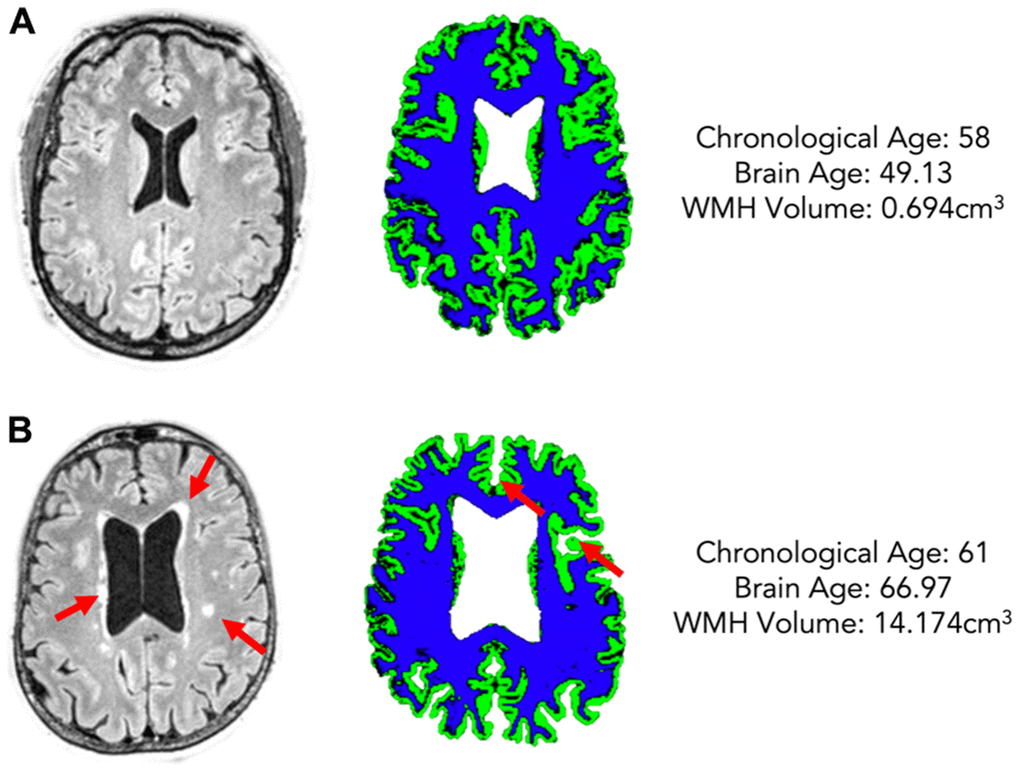
Figure 2.Two example participants of a similar chronological age (A: 58, B: 61) but different estimated brain age (A: 49.13, B: 66.97) and different WMH volume (A: 0.694cm3, B: 14.174cm3). The left column shows FLAIR scans with WMHs highlighted by red arrows, the right shows grey matter (green) and white matter (blue) maps for each participant.