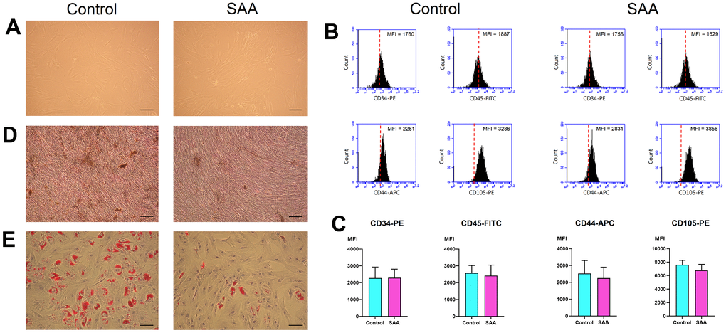
Figure 1.Characterization of MSCs. (A) In vitro culture, MSCs in the control and SAA groups shared similar growth patterns and morphologies (×100; scale bar = 100 μm). (B) Using flow cytometry, these cells were negative for CD34 and CD45, and positive for CD44 and CD105. The red dashed line indicates the isotype control; the black area indicates the stained cells. (C) There was no significant difference in the expressed MFI of CD34-PE, CD45-FITC, CD44-APC, and CD105-PE between the SAA group and the control group. (D) Osteogenic differentiation was demonstrated by mineralized deposits stainable with von Kossa stain (×100; scale bar = 100 μm). (E) Adipogenic differentiation was confirmed by intracellular accumulation of lipid droplets stainable with oil red O (×100; scale bar = 100 μm). MFI: Mean fluorescence intensity; ns: not significant; SAA: Severe aplastic anemia.