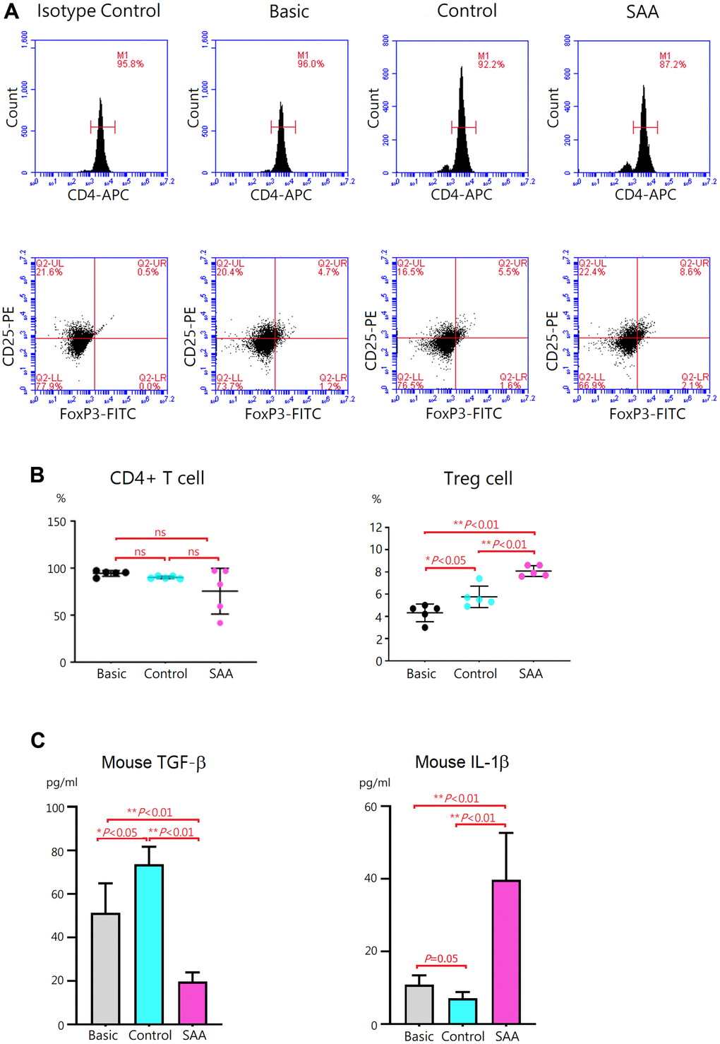
Figure 4.Evaluation of Treg differentiation after 5-days differentiation induction. (A) Differentiation of Treg cells was confirmed by intracellular staining for FoxP3 with flow cytometry. (B) The percentage of CD4+CD25+ FoxP3+ Treg cells was higher in the control group, compared to the basic group (5.8 ± 0.8% vs 4.3 ± 0.8%, P < 0.05). The percentage of CD4+CD25+ FoxP3+ Treg cells was significantly increased in the SAA group, compared to the control groups (8.1 ± 0.5% vs 5.8 ± 0.8%, P < 0.01). (C) In the SAA groups, TGF-β concentrations in the culture supernatant were decreased and IL-1β levels were increased, compared to the control group (P < 0.01). Data are presented as the mean ± SD. n = 5 in each group.