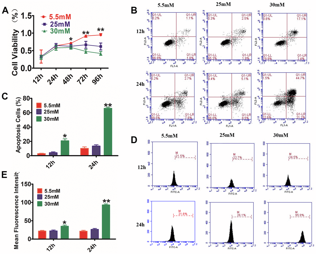
Figure 2.Glucose effects on HaCaT cells by CCK-8 assay, AnexinV-PI, and ROS staining. (A) HaCaT cells were treated with different concentrations of glucose for varying time periods, and then cell viability was measured with CCK-8. The data showed that glucose inhibited the cell viability of HaCaT cells in a dosage- and time-dependent way. (B) The annexin V-PI flow cytometry assay was utilized for the detection of the apoptosis rate of HaCaT cells, which were treated with 5.5, 25, or 30 mM glucose at different times. (C) The histogram results show that cell apoptosis increased following incubation with glucose. (D, E) Analysis of intracellular ROS levels using the flow cytometry assay. The histogram results show that the fluorescence intensity was increased following incubation with glucose. Control to 5.5 mM glucose, *P<0.05, **P<0.01, all results are representative of three separate experiments (means ± SD).