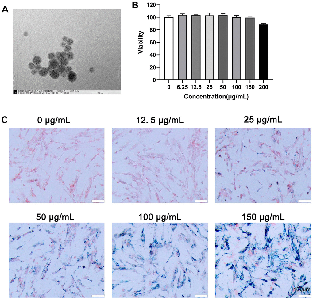
Figure 1.Characterization, viability, and internalization potential of polydopamine-capped Fe3O4 nanoparticles (MIONs@PDA). (A) Transmission electron microscopy imaging of MIONs@PDA. Scale bar = 50 nm. (B) Proliferation of human umbilical cord mesenchymal stem cells (HUMSCs) labeled with MIONs@PDA at concentrations of 0, 6.25, 12.5, 25, 50, 100, 150, and 200 μg/mL by Cell Counting Kit-8 assay. (C) Morphology of HUMSCs labeled with the MIONs@PDA at concentrations of 0, 25, 50, 75, 100, and 150 μg/mL. Scale bars = 100 μm.