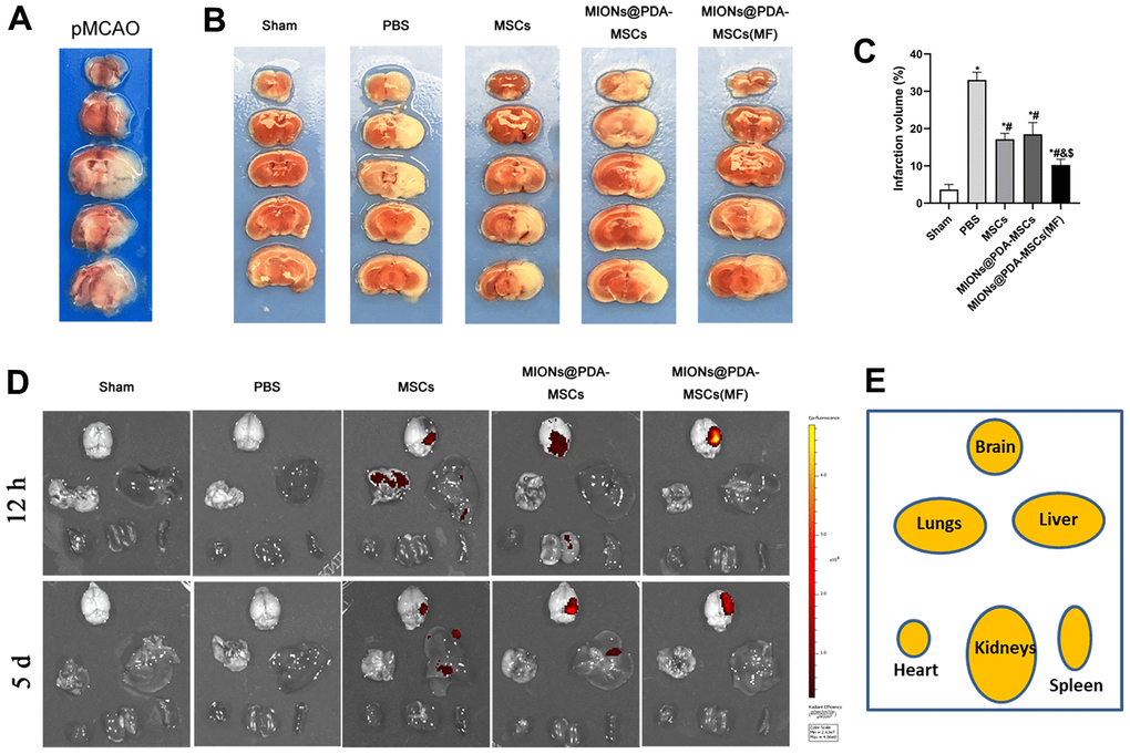
Figure 3.Effects of HUMSCs on infarct volume and behavioral improvement. (A) Representative brain slices with infarction volume shown by TTC staining. (B, C) HUMSCs treatment significantly reduced infarct volume. (D, E) Bio-distribution of the MSCs following their intravenous injection into the pMCAO-induced mice with or without the MF, evaluated by the IVIS imaging of major organs. Data are presented as the means ± standard deviation. HUMSC, human umbilical cord mesenchymal stem cell; MIONs@PDA-MSCs, HUMSCs labeled with polydopamine-capped Fe3O4 nanoparticles; MIONs@PDA-MSC(MF), MIONs@PDA-MSCs with external magnetic field; PBS, middle cerebral artery occlusion with phosphate-buffered saline administration; Sham, sham operation; TTC, 2,3,5-triphenyl-2H-tetrazoliuM chloride. *p < 0.05 vs. Sham group, #p <0 .05 vs. PBS group, &p <0 .05 vs. HUMSCs group, $p <0 .05 vs. MIONs@PDA-MSCs group.