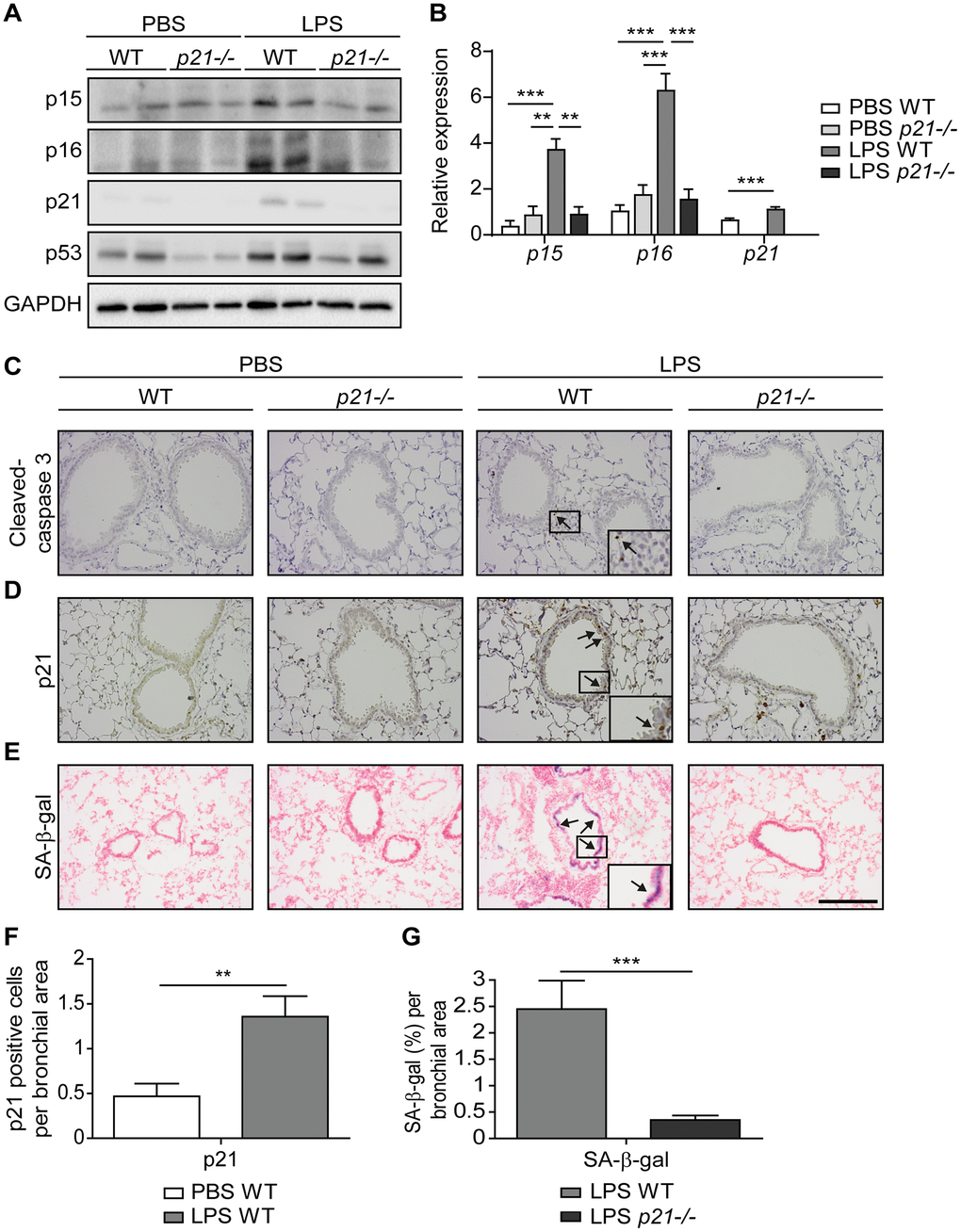
Figure 1.Accumulation of senescent cells is decreased in the lungs of p21-/- mice. WT and p21-/- mice were exposed to either PBS or aerosolized LPS (0.5 mg/ml), 3 times a week for 10 weeks. At 48 hours following the last LPS exposure, the lungs were harvested and frozen. Alternatively, lungs were harvested, fixed, and analyzed for markers of senescence. (A) Representative immunoblots for senescence-associated proteins p15, p16, p21 and p53 in the mice lungs. (B) mRNA expression levels of senescence markers p15, p16 and p21 in the mice lungs. (C, D) Immunohistochemistry (IHC) of lung sections for cleaved caspase 3, (C) and p21, (D). Scale bar represents 200μm. (E) SA-β-Gal staining of lung sections. Scale bar represents 200μm. (F) Quantification of the number of p21 positive cells per bronchial area, of the lung sections presented in (D). (G) Quantification of SA-β-gal (%) per bronchial area, of the lung sections presented in (E). Data information: Data were analyzed using one-way ANOVA, *P<0.05. **<0.005. ***P<0.0005 (B), and by Student’s t-test, *p<0.05, **p<0.01, and ***p<0.005 (F, G). Data represent mean ±SEM (A, n=3; B, n=3-10; C–G, n=3-6 independent repeats).