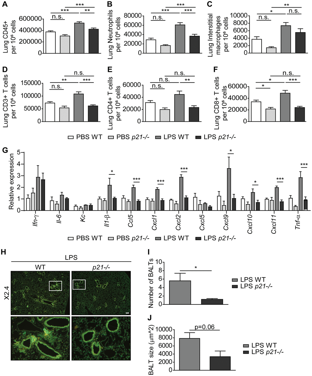
Figure 3.p21-/- decreases chronic inflammatory responses and iBALT formation caused by exposure to aerosolized LPS. WT and p21-/- mice were exposed to either PBS or aerosolized LPS (0.5 mg/ml), 3 times a week for 10 weeks. (A–F) At 48 hours following the last LPS exposure, whole lungs were dissociated into single cell suspensions and analyzed by flow cytometry to determine: (A) numbers of immune cells (CD45+), (B) numbers of neutrophils (CD45+/Ly6G+/CD11b+), (C) numbers of interstitial macrophages (CD45+/CD11c+/SiglecF-/CD11b+/CD24+), (D) numbers of CD3+ T cells (CD45+/CD3+), (E) numbers of CD4+ T cells (CD45+/CD3+/CD4+), and (F) numbers of CD8+ T cells (CD45+/CD3+/CD8+). (G) mRNA expression levels of the indicated SASP factors in the mice lungs. (H) Representative images of lungs stained for CD3+ (red) and B220 (green) depict accumulation of iBALTs in LPS exposed mice. Scale bar represents 200μm. (I, J) Numbers (I) and sizes (J) of iBALTs in the lungs of mice exposed to aerosolized LPS. Data information: Data were analyzed using one-way ANOVA, *P<0.05. **<0.005. ***P<0.0005 (A–G), and by Student’s t-test, *p<0.05, **p<0.01, and ***p<0.005 (I, J). Data represent mean ±SEM (A–F, n=9-12; G, n=3-6; H–J; n=4-6 independent repeats).