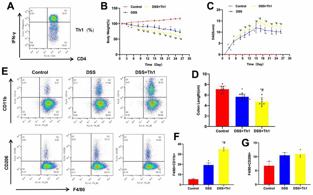
Figure 2.Th1 promoted the aggravation of mouse colitis and M1 polarization of tissue macrophages. (A) Th1 cells were highly pure after induction and amplification, which were used for subsequent experiments. (B) Dynamic detection of mouse body weight (n=10). The mouse body weight apparently declined after DSS administration, and the difference was significant compared with Control group, while Th1 injection further reduced mouse body weight. *P<0.05 compared with Control group, #P<0.05 compared with DSS group. (C) DAI scores of mice (n=10). DAI scores in DSS group significantly increased compared with Control group. In dynamic detection, the DAI scores increased with time, Th1 increased the DAI score, and the difference was significant compared with DSS group. *P<0.05 compared with Control group, #P<0.05 compared with DSS group. (D) Mouse intestinal length (n=10). Relative to Control, the intestinal tissue length of DSS mice was shortened, Th1 injection aggravated mouse intestinal injury, and further reduced the intestinal tissue length. *P<0.05 compared with Control group, #P<0.05 compared with DSS group. (E–G) Proportions of M1/M2 macrophages (n=10). The proportion of M1 cells in DSS increased, and the difference was significant compared with Control group. The proportion of M1 cells in DSS+Th1 group further increased, higher than that in DSS group. There was no significant difference in M2 cell proportion. *P<0.05 compared with Control group, #P<0.05 compared with DSS group.