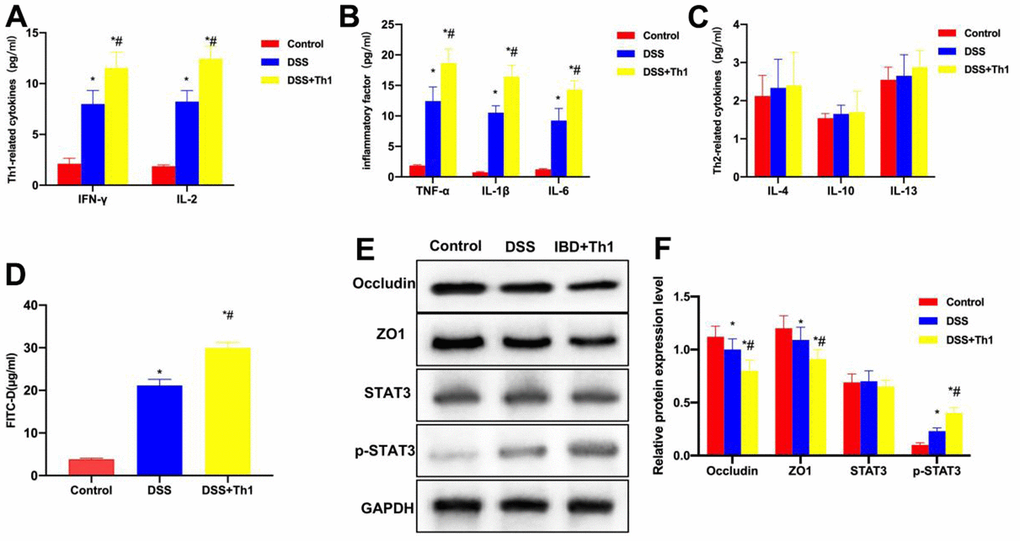
Figure 3.Th1 aggravated intestinal inflammation and reduced the TJ protein levels. (A) Th1 cell characteristic factors (n=10). The expression of IFN-γ and IL-2 in DSS significantly increased, while that in DSS+Th1 group further increased relative to DSS group, and the difference was significant compared with DSS group. *P<0.05 compared with Control group, #P<0.05 compared with DSS group. (B) Detection of inflammation cytokines (n=10). The levels of TNF-α, IL-6 and IL-1β in DSS group significantly increased, higher than those in Control group. The inflammatory factor levels in DSS+Th1 group further increased, and Th1 improved their tissue levels. *P<0.05 compared with Control group, #P<0.05 compared with DSS group. (C) Th2 cell characteristic factors (n=10). The levels of IL-4, IL-10 and IL-13 were not significantly different among Control, DSS and DSS+Th1 groups. (D) FITC-D (n=10). The FITC-D penetration rate in DSS group significantly increased, while that in DSS+Th1 group further increased relative to DSS group, and the mucosal permeability was apparently enhanced. *P<0.05 compared with Control group, #P<0.05 compared with DSS group. (E, F) Protein detection results (n=5). DSS induced the down-regulation of TJ proteins ZO1 and Occuldin, and their expression further decreased in DSS+Th1 group. At the same time, the STAT3 phosphorylation level increased. *P<0.05 compared with Control group, #P<0.05 compared with DSS group.