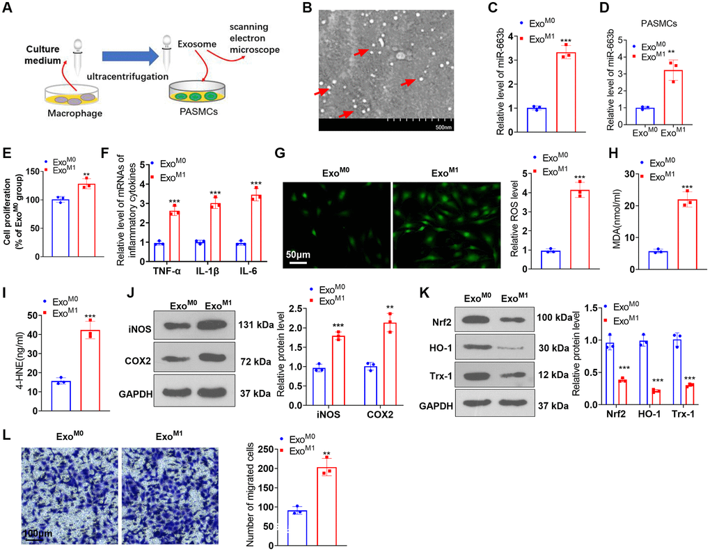
Figure 4.miR-663b presented a high level in M1 macrophage exosomes, and M1 macrophage exosomes elicited PASMC damage. (A) The exosomes of M0 and M1 macrophages were acquired through ultracentrifugation. (B) The isolated exosomes were identified by scanning electron microscope (SEM). Scale bar = 500 nm. (C) RT-PCR was carried out to check miR-663b expression in M0 and M1 macrophage exosomes. M1 macrophage exosomes were cultured together with PASMCs. (D) RT-PCR was carried out to check miR-663b expression in PASMCs. (E) CCK8 assay was conducted for examining PASMC proliferation. (F) RT-PCR checked the profiles of inflammatory factors in PASMCs. (G–I) Cell immunofluorescence and colorimetry determined the levels of ROS, MDA, and 4-HNE in PASMCs. (J, K) Western blot confirmed the profiles of inflammation-concerned proteins and oxidative stress-associated proteins in PASMCs. (L) Transwell measured PASMC migration. N = 3. *P < 0.05, **P < 0.01, ***P < 0.001 (vs. ExoM0).