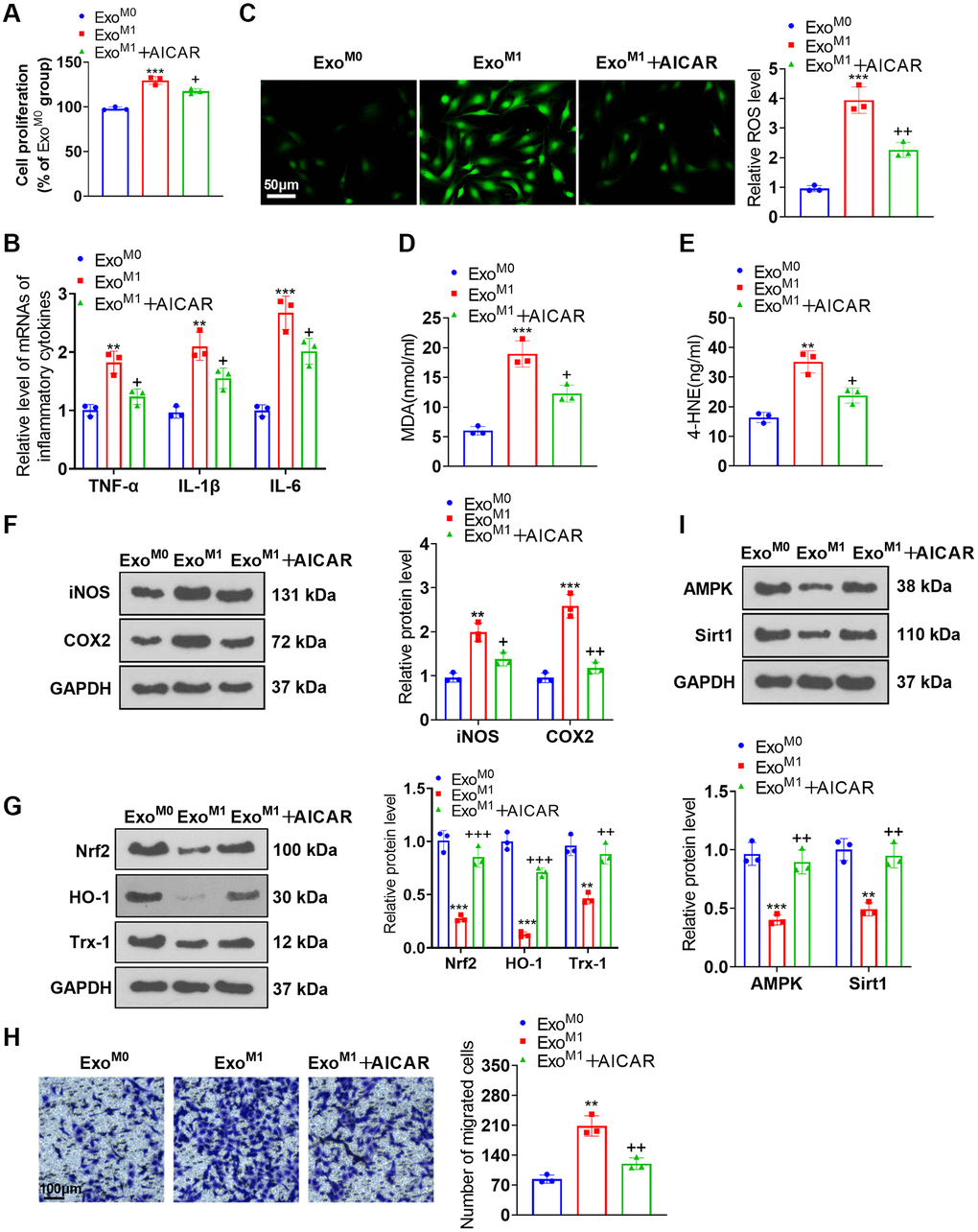
Figure 7.AMPK activation ameliorated the impacts of M1 macrophage exosomes on PASMCs. PASMCs were cultured together with M0 and M1 macrophage exosomes, with AICAR applied to the conditioned medium. (A) CCK8 assay was conducted for examining PASMC proliferation. (B) RT-PCR was taken for gauging TNF-α, IL-1β, and IL-6 expressions in PASMCs. (C–E) Cell immunofluorescence and colorimetry confirmed the levels of ROS, MDA, and 4-HNE in PASMCs. (F, G) Western blot was done for measuring iNOS, COX2, Nrf2, HO-1 and Trx-1 expressions in PASMCs. (H) Transwell examined PASMC migration. (I) Western blot verified the level of the AMPK/Sirt1 pathway in PASMCs. N = 3. **P < 0.01, ***P < 0.001 (vs. ExoM0); +P < 0.05, ++P < 0.01, +++P < 0.001 (vs. ExoM1).