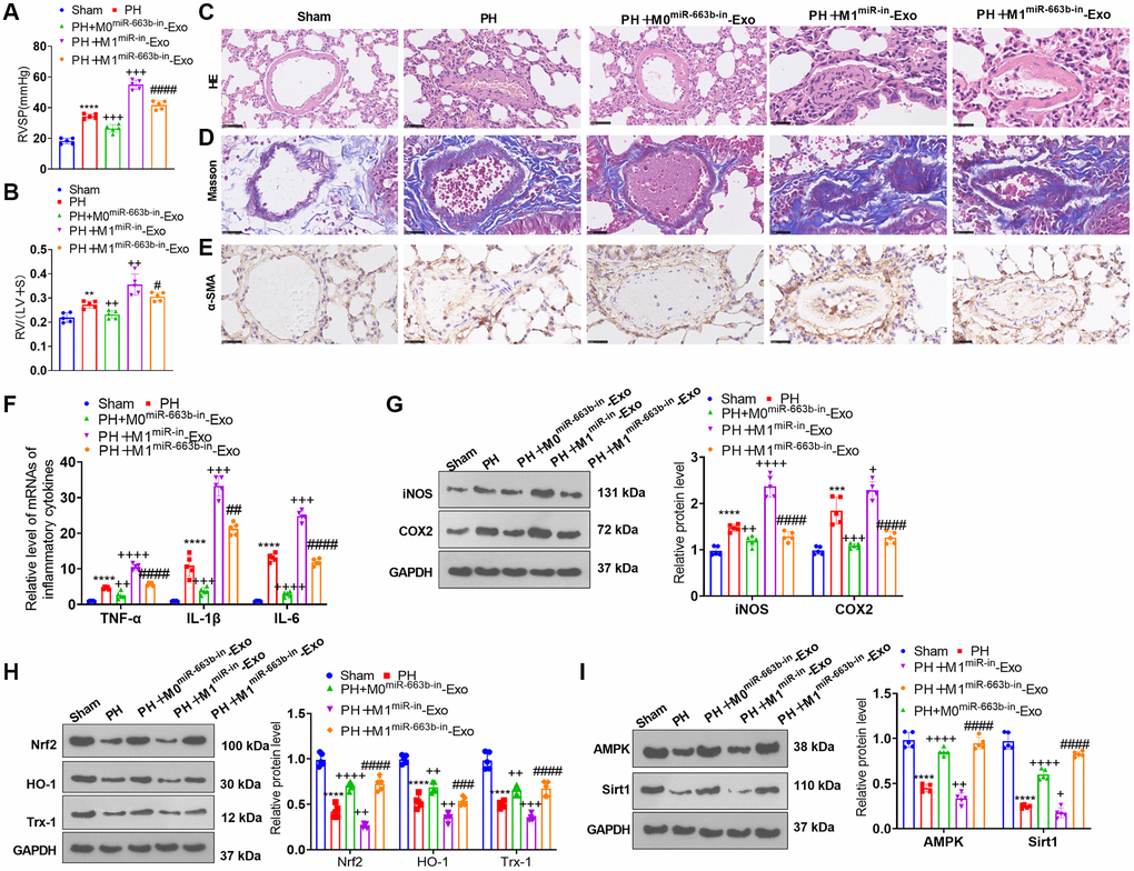
Figure 8.M1 macrophage exosomes with miR-663b low expression ameliorated pulmonary vascular remodeling in pulmonary hypertension rats. A pulmonary hypertension rat model elicited by hypoxia was built, and M1 macrophage exosomes with low-expressed miR-663b were transfused into the caudal veins of the rats. (A, B) The hemodynamic assay was carried out to confirm the mean ratio of RVSP and RV/(LV+S) of PH rats. (C, D) HE staining (C) and Masson staining (D) were conducted for monitoring pathological alterations in the lung tissues of PH rats. (E) Immunohistochemistry (anti-α-SMA) was performed for detecting pulmonary vascular remodeling. (F) RT-PCR was implemented for gauging TNF-α, IL-1β, and IL-6 levels in the tissues. (G, H) iNOS, COX2, Nrf2, HO-1 and Trx-1 profiles in the tissues were confirmed through western blot. (I) The level of the AMPK/Sirt1 pathway in the tissues was ascertained by western blot. N = 5. **P < 0.01, ***P < 0.001, ****P < 0.0001 (vs. Sham); ++++P < 0.0001 (vs. PH); ####P < 0.0001 (vs. PH+M1miR-in-Exo).