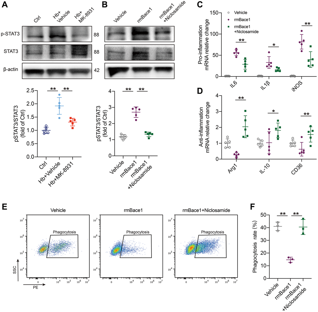
Figure 5.BACE1 induced microglia pro-inflammatory phenotype by STAT3 signaling activation. (A) The levels of phosphorylated and total STAT3 relative to the internal control actin are shown in the indicated treated PMG. n = 5 per group. (B) Primary microglia were treated with rmBACE1 protein with/without niclosamide treatment. Phosphorylated and total STAT3 were determined 12 h after treatment. n = 5 per group. (C, D) Quantification of pro- or anti-inflammatory cytokine gene expression is shown in the bar graphs, with data from at least 5 indicated treated PMGs. Data are shown as the relative change compared to the vehicle group. (E, F) In vitro analysis of the PMG erythrophagocytosis rate under rmBACE1 stimulation with/without niclosamide treatment, n = 3 per group. All data are presented as the mean ± SD. *P < 0.05, **P < 0.01.