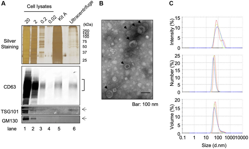
Figure 1.Isolation of EVS from cultured keratinocytes. Conditioned media from human primary keratinocytes were collected. EVs were isolated from these media by an ultracentrifugation protocol. (A) The expression of specific markers for EVs (CD63 and TSG101) were observed in the ultracentrifugation purified EVs fraction (Ultracentrifuge Lane), whereas the expression of GM130 were not observed. 20 μg, 2 μg, 0.2 and 0.02 μg of total cell lysates were also analyzed as well as an EV fraction purified by the Qiagen exoEasy kit (Kit A lane) (B) Analysis of ultracentrifuge purified EVs fraction by transmission electronic microscopy. (C) Hydrodynamic size distribution profiles of ultracentrifuge purified EVs were evaluated by DLS, using either intensity, number or volume quantification. The three color curves correspond to three series of 10 measurements (technical replicates). All experiments were repeated independently at least three times (biological replicates) and a representative result is shown.