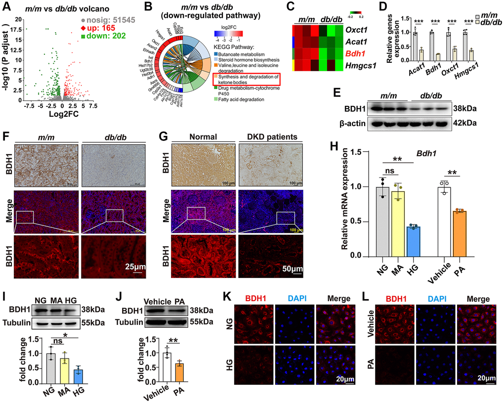
Figure 1.Expression of BDH1 is downregulated in diabetic kidney and HG- or PA-treated HK-2 cells. (A) Volcano plot showing differentially expressed genes (DEGs) (red, upregulated genes; green, downregulated genes) in the kidneys of m/m and db/db mice (n = 3 mice per group). (B) KEGG enrichment analysis showing the top 6 downregulated pathways. (C) Four DEGs involved in ketone body synthesis and degradation pathways. (D) qRT-PCR analysis showing the mRNA levels of Acat1, Bdh1, Oxct1, and Hmgcs1 in the kidneys of m/m and db/db mice (n = 3 mice per group). WB (E), IHC, and IF (F) showing the protein level of BDH1 in the kidneys of m/m and db/db mice. IHC and IF (G) showing the protein level of BDH1 in the kidneys of normal subjects (n = 9) and patients with DKD (n = 8). (H) qRT-PCR showing the mRNA level of Bdh1 in HK-2 cells treated with HG or PA. Representative WB images showing the protein level of BDH1 in HK-2 cells stimulated with HG (I) or PA (J). Representative IF images showing the protein level of BDH1 in HK-2 cells stimulated with HG (K) or PA (L). All results are representative of three independent experiments. Values are presented as mean ± standard deviation. Bar: 50 and 25 μm in F, and 100 and 50 μm in G, 20 μm in K and L. Abbreviations: BDH1: β-hydroxybutyrate dehydrogenase 1; NG: normal glucose; HG: high glucose; PA: palmitic acid; MA: mannitol; DEGs: differentially expressed genes; WB: western blot; IHC: immunohistochemistry; IF: immunofluorescence; DKD: diabetic kidney disease. *P < 0.05; **P < 0.01; ***P < 0.001; Abbreviation: ns: no significance.