Submit an Article
Navigate
Home
Editorial Board
Editorial Policies
Current Volume
Archive
Scientific Integrity
Publication Ethics Statements
Interviews with Outstanding Authors
Newsroom
Sponsored Conferences
Podcast
Contact
Special Collections
Submit an Article
Online ISSN: 1945-4589
Research Paper
|
Volume 15, Issue 23
|
pp. 14292–14305
Delivering umbilical cord mesenchymal stem cell exosomes through hydrogel ameliorates vaginal atrophy in ovariectomized rats
Back to article
Figure 6
(6 of 6)
−
100%
+
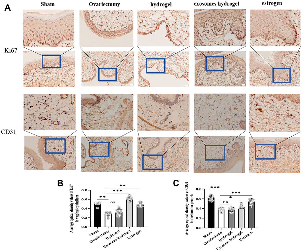
Figure 6.
IHC analysis.
(
A
) Average optical density analysis of ki67 and CD31 expression in the vaginal epithelium and lamina propria; (
B
,
C
) Quantification of ki67 and CD31 staining.