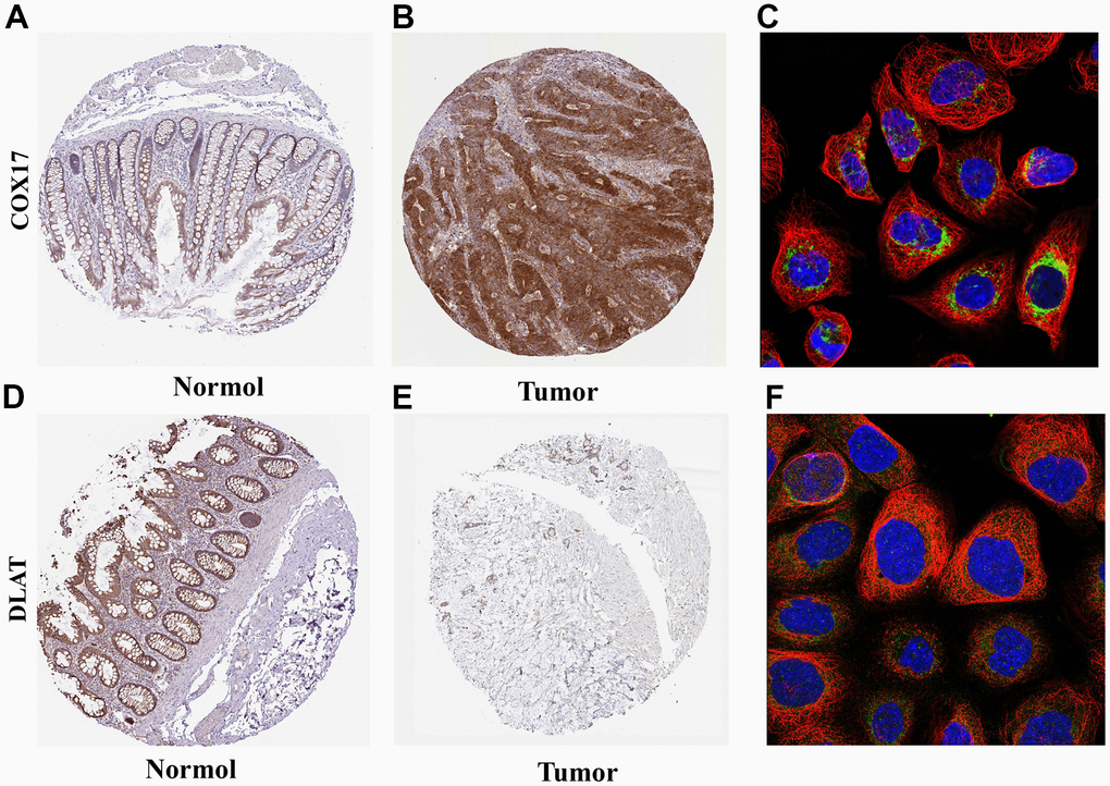
Figure 7.Expression and subcellular localization of COX17 and DLAT (A) The expression of COX17 is greater in tumor tissues compared to (B) normal tissues. (C) COX17 was mainly located in mitochondria. DLAT expression in (D) CRC and (E) normal issues. (F) DLAT were mainly located in the mitochondria by the HPA database analysis.