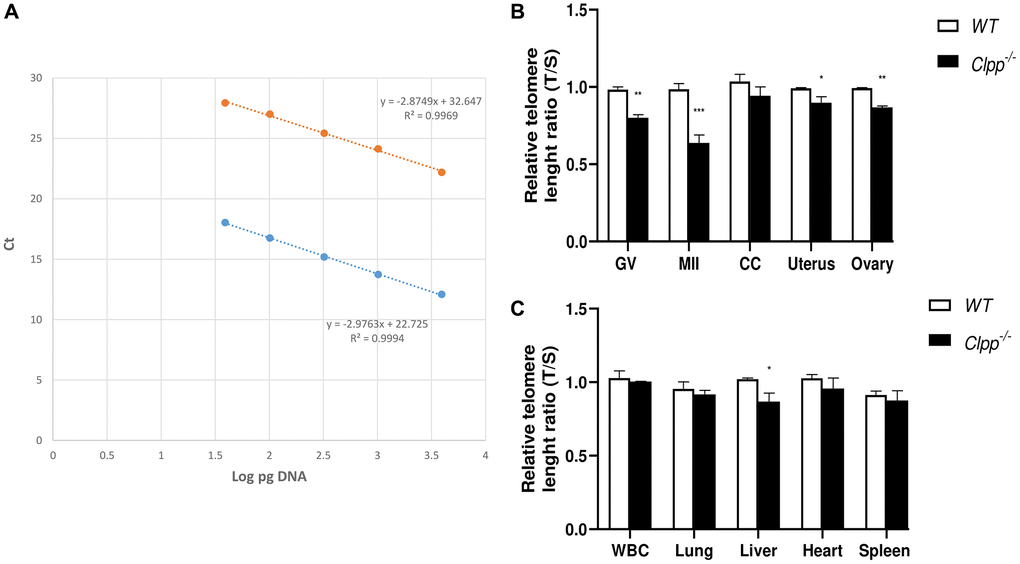
Figure 2.Telomere length in oocytes, ovaries, and somatic tissues of 6-month-old Clpp−/− and WT mice. (A) The standard curve was generated by serial dilution of known amounts of DNA to calculate relative DNA concentrations (log DNA) from Ct values of the qPCR products. Blue dots: telomere; orange dots: 36B4 single copy gene (control). The correlation regression equation and coefficients (R2) of Ct versus log DNA are shown. (B, C) The relative telomere lengths of GV and MII oocytes, ovaries and somatic cells and tissues are represented as ratios of T/S. Abbreviations: GV: Germinal vesicle, CC: Cumulus cells, WBC: White blood cells. Data presented as mean ± SD. **p < 0.01, *p < 0.05 using t-test. The telomere length assessment was repeated twice, using five mice (ten GVs from each) in each group per experiment.