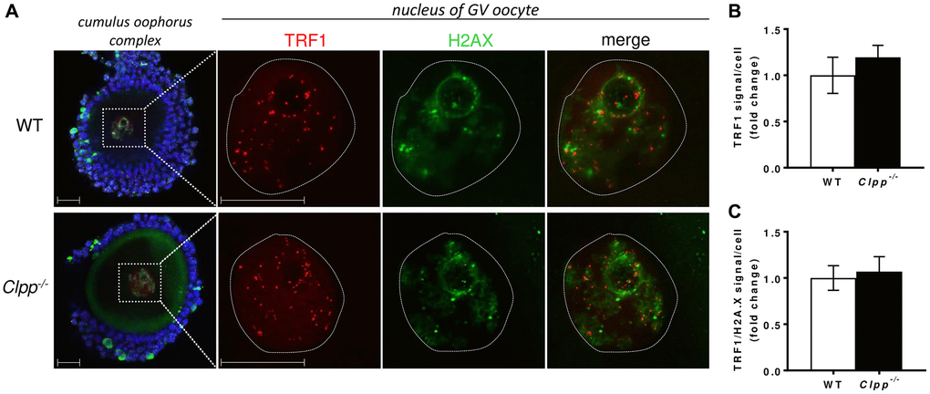
Figure 5.Representative confocal images of TRF1 expression and TRF/H2AX co-localization in cumulus oophorus isolated from 2-month-old wild-type and Clpp−/− mice. (A) Immunofluorescence double staining of TRF1 (red) and H2AX (green) in cumulus oophorus complexes of 2-month-old WT and Clpp−/− mice. Nuclear area of GV oocytes is highlighted by a dotted line. Nuclei were stained for TRF1 (red) and H2AX (green). DAPI (Blue) was used to stain nuclei (blue). The highlighted box in Clpp−/− sample shows co-localization of TRF1 and H2AX (white arrow). Scale bar = 25 μm. DAPI was used to stain nuclei (blue). (B) Quantitative analysis of TRF1 immunofluorescence in WT and Clpp−/− GV oocytes. (C) Quantitative analysis of co-localization of TRF1 and H2AX in WT and Clpp−/− GV oocytes. Data presented as mean ± SD with t-test (**p < 0.01, *p < 0.05). Experiments repeated twice, using three mice (five GVs from each) in each group per experiment.