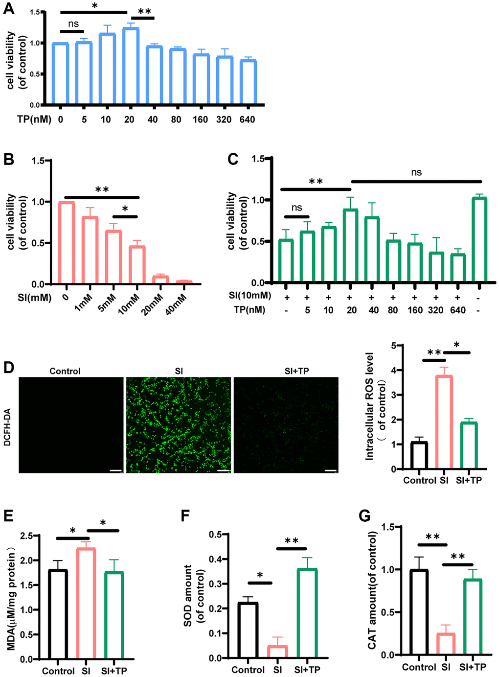
Figure 1.The effects of triptolide on the levels of pro- and anti-oxidant factors and the cell viability. The ARPE-19 cells were incubated with different concentrations of triptolide (TP) (A) or sodium iodate (SI) (B) for 24 h. (C) The ARPE-19 cells were treated with TP for different concentrations and then exposed to 10 mM SI for 24 h, n = 4. TP prevented the decrease in retinal pigment epithelial cell viability induced by SI. The fluorescence images of ROS were measured by a fluorescence microplate, n = 3. (D) The data showed that TP reduced the generation of ROS in ARPE-19 cells significantly. The amounts of MDA (E), SOD (F), CAT (G) in cell lysates were detected by a microplate reader using commercial kits. Data are shown as mean ± standard deviation (SD), n = 3; Scale bar, 300 μm. In all cases, the control is untreated retinal pigment epithelial (RPE) cells. Abbreviation: NS: not significant. *P < 0.05. **P < 0.01.