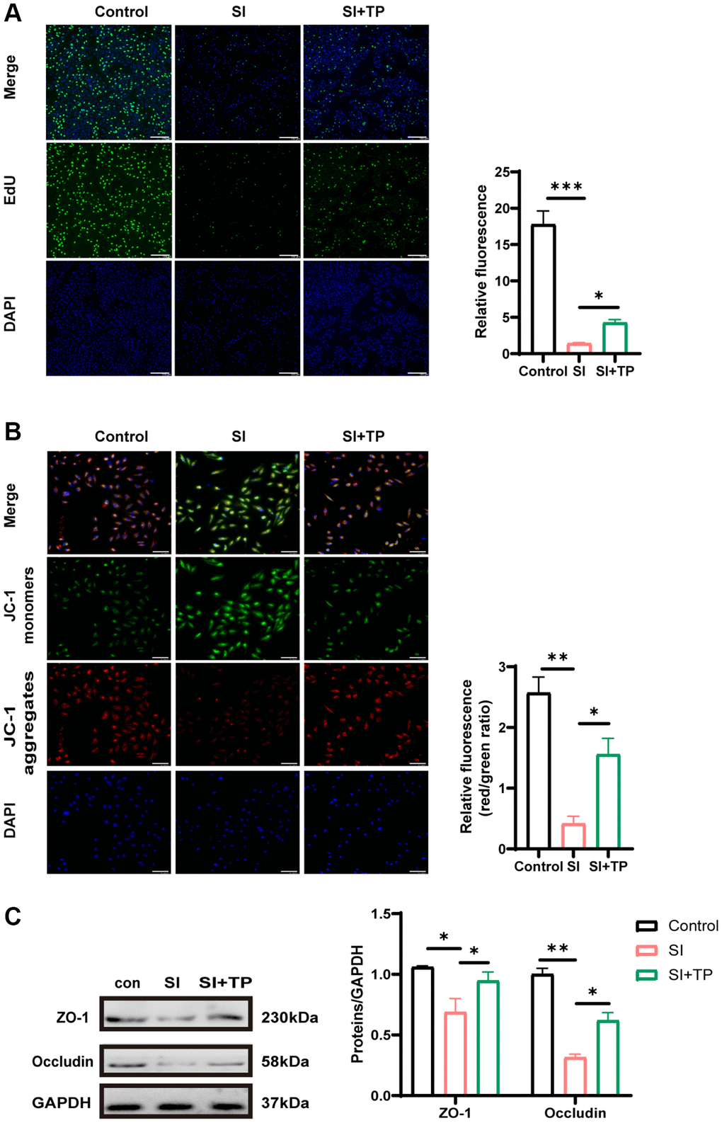
Figure 2.Effects of TP on proliferation, mitochondrial membrane potential and tight junction protein. (A) The EdU assay was used to analyze the proliferation-suppressing effect of SI on RPE cells. DAPI for nuclear staining (blue). EdU-positive cells (green) were counted to calculate the percentage. Scale bar, 200 μm. (B) Mitochondrial membrane potential was detected by the JC-1 fluorescence ratio. The transition from red fluorescence to green fluorescence represents the decrease of cell membrane potential. Scale bar, 200 μm. (C) Protein levels of occludin and ZO-1 were detected by Western blot with GAPDH as the loading control. The bar graphs show the results of analysis. Values are the mean ± SD; n = 3; *P < 0.05, **P < 0.01, ***P < 0.001.