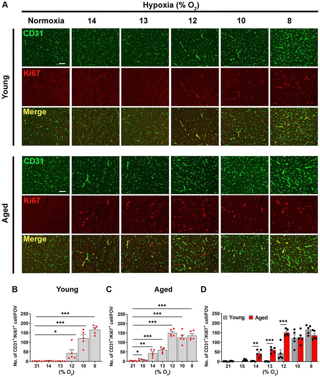
Figure 1.Hypoxia-induced cerebrovascular remodeling is triggered at a higher O2 level in aged mice. (A) Frozen brain sections taken from young (2 months) and aged (20 months) mice exposed to normoxia or different levels of hypoxia (15–8% O2) for 4 days were stained for the endothelial marker CD31 (AlexaFluor-488) and Ki67 (Cy-3). Images were captured in the midbrain. Scale bars = 100 μm. (B–D) Quantification of the density of CD31+/Ki67+ cells following normoxia or 4 days exposure to the different levels of hypoxia in young (B), aged (C) and both combined (D). Results are expressed as the mean ± standard error of the mean (SEM) (n = 5 mice/group). *p < 0.05, **p < 0.01, ***p < 0.001. One-way analysis of variance (ANOVA) followed by Tukey’s multiple comparison post-hoc test. Note that aged mice are more sensitive to the effects of hypoxia in that they display endothelial proliferation at a higher O2 level than young mice.