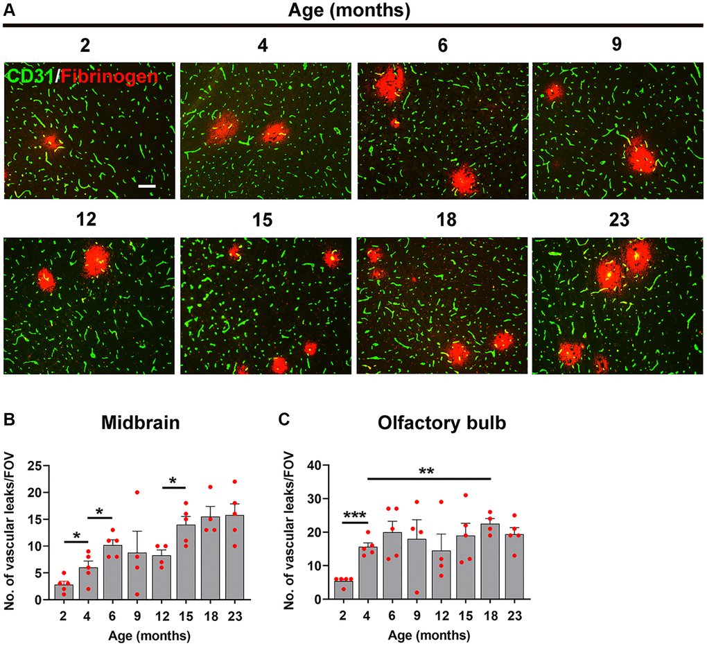
Figure 4.Defining the age at which the BBB becomes more susceptible to hypoxia. (A) Frozen brain sections taken from mice of different ages (2–23 months) exposed to hypoxia (8% O2) for 4 days were dual-stained for CD31 (AlexaFluor-488) and fibrinogen (Cy-3). Images were captured in the midbrain. Scale bars = 100 μm. Quantification of the density of extravascular leaks in the midbrain (B) or olfactory bulb (C) in different age mice following 4 days hypoxia. Results are expressed as the mean ± standard error of the mean (SEM) (n = 5 mice/group). *p < 0.05, **p < 0.01, ***p < 0.001. One-way analysis of variance (ANOVA) followed by Tukey’s multiple comparison post-hoc test. Note that the density of vascular leaks increased with age and that major increases were seen between 2–6 months, as well as between 12–15 months in the midbrain.