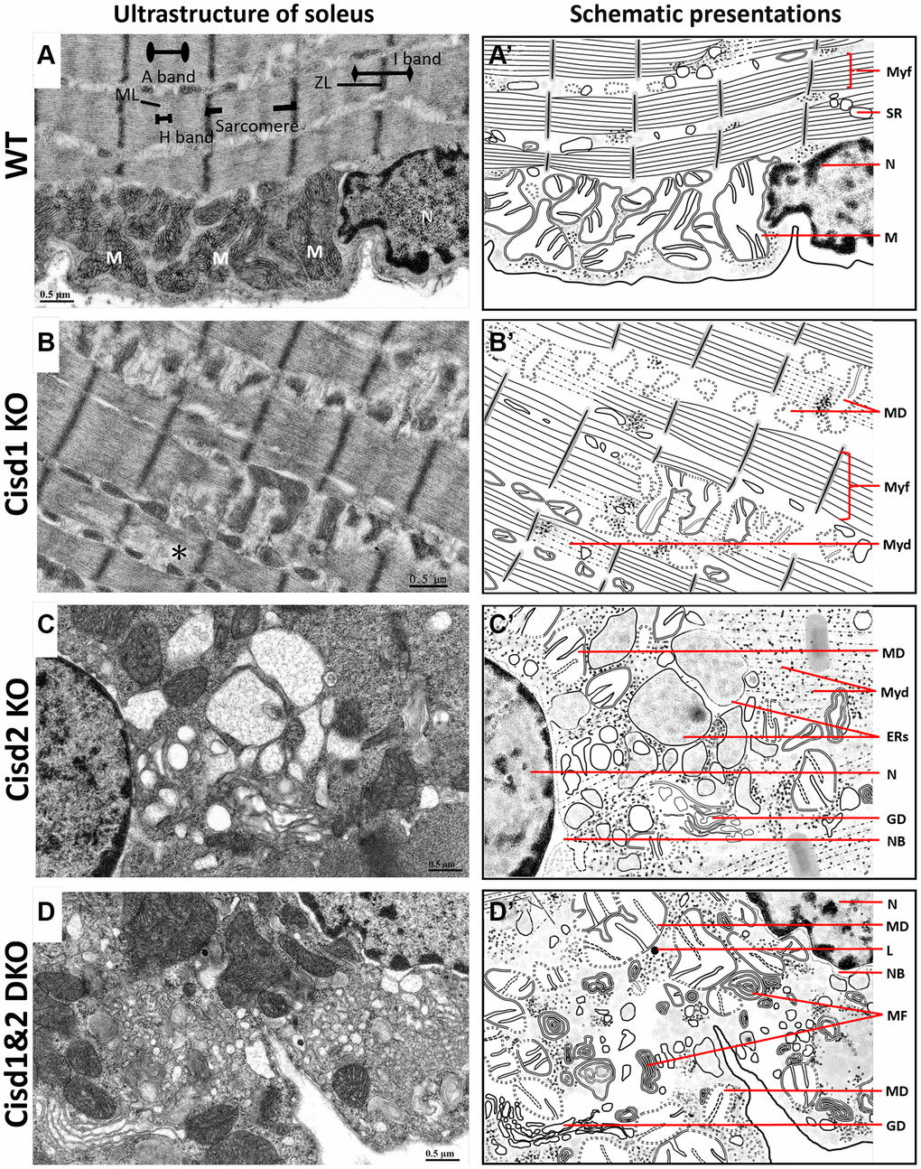
Figure 4.Ultrastructure of skeletal muscle (soleus) from Cisd1&2 DKO mice. (A) Architecture of the soleus in WT mice. ZL, Z line; ML, M line; M, mitochondria; N, nucleus. (B) Mitochondrial defects, myofibril degeneration (*) and ER stress in Cisd1 KO soleus. (C) Mitochondrial defect, myofibril degeneration, necrosis and ER stress in Cisd2 KO soleus. (D) Mitochondrial defect, myofibril degeneration, necrosis and ER stress in Cisd1&2 DKO soleus. (A–D) were shown at higher magnification (yellow squares) in Supplementary Figure 3. (A’–D’) are schematic presentations of the ultrastructure of the soleus shown in (A–D). Abbreviations: Ne: Nucleolus; NB: Nuclear envelope breakdown; MD: Mitochondrial defect; Myf: Myofibril; Myd: Myofibril degeneration; Ers: ER stress; ER: Endoplasmic Reticulum; SR: Sarcoplasmic Reticulum; Avi: Autophagosome; Avd: Autolysosome; L: Lysosome; MF: Myelin Figure; GD: Golgi degeneration. Mouse age, 5 weeks old.
Figure 4 — Cisd1 synergizes with Cisd2 to modulate protein processing by maintaining mitochondrial and ER homeostasis | Aging