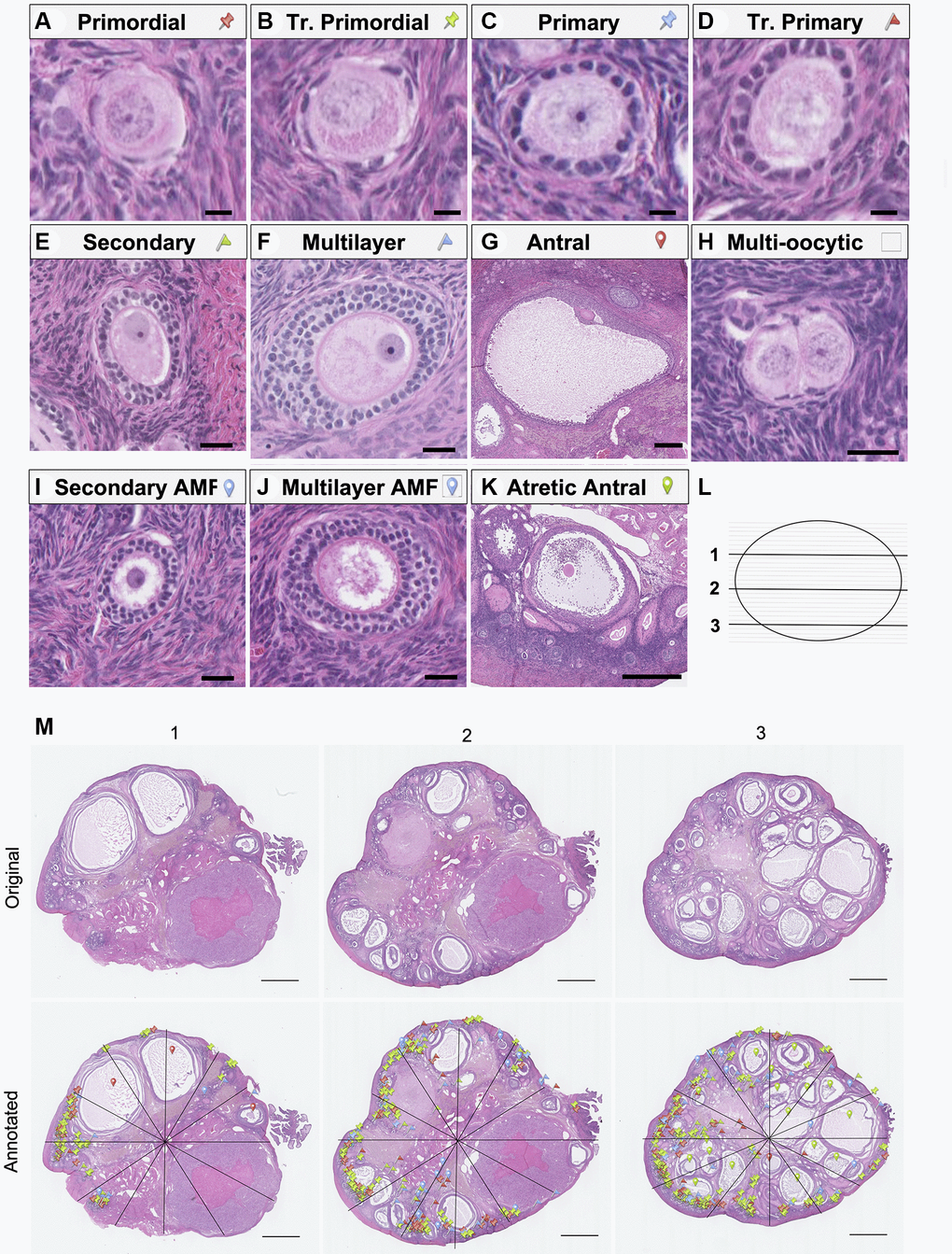
Figure 3.Representative images of follicle classes and follicle counting method schematic. Representative hematoxylin-and-eosin-stained images illustrating the following follicle classes are shown: (A) primordial, (B) transitional primordial), (C) primary, (D) transitional primary, (E) secondary, (F) multilayer, (G) antral, (H) multi-oocytic, (I) secondary with abnormal follicle morphology (AMF), (J) multilayer with AMF, (K) and atretic antral follicles. The follicle counting workflow depicting (L) the selection of representative ovarian sections from the top (1), middle (2), and bottom (3) quartiles of the ovary, and (M) schematic of follicle counting workflow, including dividing each section into twelve radial segments and annotation with colored markers as defined in A-K to mark and classify each follicle class. The scale bars are 10 μm (A–D), 25 μm (E, F, H, I and J), 250 μm (G), 500 μm (K), and 1 mm (N).