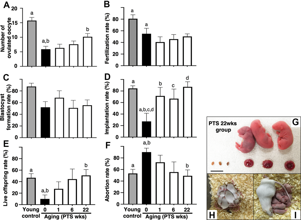
Figure 2.Effects of pterostilbene ingestion on reproductive outcomes in aged mice. Ovulation was induced at the proestruus stage following 47 weeks of pterostilbene (PTS) ingestion by administrating human chorionic gonadotropin (hCG). At 15 hours post-hCG injection, cumulus-oocyte complexes (COCs) were collected from the oviduct ampulla and subsequently inseminated with sperm from fertile male mice. At 16 hours after culture, 2-cell stage embryos were collected and allowed to develop to the blastocyst stage for an additional 72 hours of culture. Following the completion of embryo culture, the blastocysts from each animal were transferred to independent recipient mice. A Caesarian section was performed 16 days after embryo transfer to ascertain the number of implantation sites and live offspring. For the young control group, ICR mice at six weeks of age were used (young control). (A) Number of ovulated oocytes. The number of ovulated oocytes was quantified by the removal of cumulus cells surrounding oocytes after insemination using a stereomicroscope (n=18-20 animals). (B) Fertilization rate (the number of 2-cell stage embryos divided by the number of ovulated oocytes) (n=11* - 17 animals, 40-86 two-cell stage embryos per group). *, three mice in the control group, seven mice in the PTS 1-week group, four mice in the PTS 6-weeks group, and one mouse in the PTS 22-weeks group did not ovulate. (C) Blastocyst formation rate (the number of blastocysts divided by the number of 2-cell stage embryos) (n=10*-15 animals, 18-51 blastocysts per group). *, oocytes retrieved from three mice in the control group, one mouse in the PTS 1-week group, two mice in the PTS 6-weeks group, and one mouse in the PTS 22-weeks group did not fertilize. (D) Implantation rate (the number of implanted blastocysts divided by the number of transferred blastocysts) (n=9-13* animals, 10-46 implanted blastocysts per group). *, 2-cell stage embryos derived from three mice in the control group, one mouse in the PTS 1-week group, four mice in the PTS 6-weeks group, and two mice in the PTS 22-weeks group were arrested in their development before reaching the blastocyst stage. (E) Live offspring rate (the number of live offspring divided by the number of transferred blastocysts) (n=9-13 animals, 5-32 live offspring per group). (F) Abortion rate (1 minus the live offspring rate). (G) Representative images of live offspring and placentas derived from the PTS 22-weeks group. Scale bars, 10 mm. Following the Caesarian section, the offspring were nursed by foster mothers to assess their health status and were mated at 8 weeks of age to confirm their fertility. (H) The offspring at 10 days after Caesarian section. (I) The offspring with delivered pups. The bars represent the mean ± SE. Different letters (A–D) show significant differences (p < 0.05) between the same symbols.