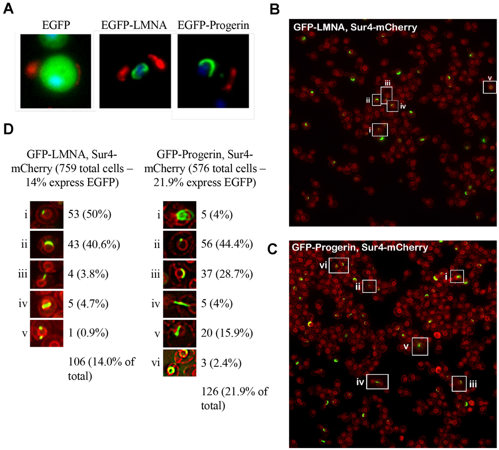
Figure 2.EGFP-LMNA and EGFP-Progerin localize to the yeast nuclear membrane. (A) Aliquots of cells from each EGFP (left panel), EGFP-LMNA (middle panel) and EGFP-Progerin (right panel) were fixed and prepared for fluorescence microscopy. Green signal indicates EGFP signal. Cells were labelled with wheat germ agglutinin (red) to mark bud scars on the cell periphery. Chromatin is counterstained with Hoechst 33342 dye to mark nuclei (blue). (B) and (C) Live cell imaging of fluorescence tags was performed in a yeast strain containing an endogenous SUR4-mCherry epitope to fluorescently mark the nuclear membrane (red). The SUR4-mCherry cells were transformed with EGFP-LMNA (B) or EGFP-Progerin (C), grown overnight in Ura- media supplemented with 2% Raf for 24h, diluted to an OD600 of 0.2 in fresh Ura- media supplemented with 2% galactose, then incubated at 30° C for 6 hours. (D) Examples of cells highlighted in (B) and (C) were shown in the inset regions, and counted. The percent of total cells expressing EGFP with different localization patterns is given.