





Introduction
Metabolic dysfunction-associated steatotic liver disease (MASLD) is a highly prevalent condition with a substantially increasing incidence in recent years. MASLD includes a spectrum of liver disorders, from simple steatosis to metabolic dysfunction-associated steatohepatitis (MASH). MASH is characterised by hepatocellular injury, specifically ballooning degeneration, and inflammation. This condition can progress to fibrosis, cirrhosis, and hepatocellular carcinoma (HCC) [1]. MASLD is increasingly recognised as the hepatic manifestation of metabolic syndrome and is closely associated with insulin resistance and systemic metabolic dysfunction [2, 3]. In addition to hepatic complications, MASLD increases the risk of extrahepatic conditions, particularly cardiovascular disease, which represents the leading cause of mortality in this population.
Lipocalin-2 (LCN2), also known as neutrophil gelatinase-associated lipocalin (NGAL), is a protein involved in lipid transport and immune responses through iron binding. LCN2 also participates in apoptosis [4], lipid metabolism [5], cell migration [6], and the regulation of receptor trafficking.
Recent studies indicate that LCN2 levels are elevated in patients with MASH and correlate with key indicators of disease severity, including inflammation, insulin resistance, and fibrosis. These associations suggest that LCN2 may serve as a biomarker for disease progression [7].
Beyond its metabolic functions, LCN2 is involved in neuroinflammatory processes. Its expression increases in the central nervous system during inflammation, where it is produced by microglia and endothelial cells and contributes to neuroimmune signalling [8–11]. LCN2 has also been implicated in immune dysregulation associated with neurodegenerative diseases, although its precise role remains unclear [12, 13].
Due to its involvement in metabolic, inflammatory, and neurovascular pathways, LCN2 may represent a potential link between MASLD and cardiocerebrovascular disease. Previous findings from this group demonstrated that patients with MASLD exhibit increased arterial stiffness, endothelial dysfunction, and reduced Mini-Mental State Examination (MMSE) scores, which suggest early cognitive impairment [14]. Additionally, greater severity of liver fibrosis has been associated with a higher risk of subclinical brain lesions, further supporting the hypothesis of systemic vascular involvement in MASLD [15].
Cardiac dysfunction in MASLD has been evaluated using myocardial mechano-energetic efficiency (MMEE), a parameter associated with insulin resistance, obesity, and type 2 diabetes mellitus, and predictive of cardiovascular events [16, 17]: evidence indicates that insulin resistance may mediate the relationship between MASLD and impaired MMEE [18, 19].
Despite these findings, no studies have yet evaluated the association between circulating LCN2 levels and surrogate markers of cardiovascular risk in patients with MASLD.
Therefore, the present study aimed to evaluate serum LCN2 levels in patients with MASLD and to determine whether elevated LCN2 levels are specifically associated with endothelial dysfunction, vascular structural changes, and impaired myocardial mechano-energetic efficiency. It was hypothesised that higher LCN2 levels would indicate increased cardiovascular risk by reflecting an integrated adipo-neurovascular inflammatory mechanism in MASLD.
Aims of the Study
The primary aim of this cross-sectional study was to evaluate the associations between adipo-neuroinflammation markers (such as LCN2), indicators of cognitive impairment (MMSE score), and surrogate cardiovascular risk indicators (RHI, IMT, and MMEE) in a cohort of MASLD patients compared with controls.
Materials and Methods
Study population
Between June 2023 and April 2024, we consecutively enrolled patients with MASLD admitted to the Gastrointestinal and Liver Unit and the Internal Medicine with Stroke Care Ward of Policlinico “Paolo Giaccone” in Palermo.
Inclusion criteria were:
• histological diagnosis of MASLD based on liver biopsy performed within 6 months prior to enrolment;
• alcohol consumption < 20 g/day in the previous year (assessed by questionnaire);
• evidence of hepatic steatosis on ultrasound and liver stiffness > 6 kPa.
Exclusion criteria included:
• liver cirrhosis;
• hepatocellular carcinoma;
• other causes of liver disease (alcoholic, viral, autoimmune, Wilson’s disease, haemochromatosis, α1-antitrypsin deficiency);
• human immunodeficiency virus (HIV) infection;
• use of steatosis-inducing drugs (steroids, amiodarone, tamoxifen);
• malignancies;
• rheumatic diseases;
• haematological disorders.
The control group included subjects without a history of cardiovascular or cerebrovascular disease (e.g., myocardial infarction, ischemic stroke), without acute illness at enrolment, and with anthropometric and metabolic characteristics comparable to those of MASLD patients, except for the absence of liver disease. Patients and controls were matched for age (±3 years) and sex. All participants provided written informed consent, and the study was conducted in accordance with the Declaration of Helsinki (2001). Clinical and anthropometric data were collected at enrolment. Body mass index (BMI) was classified as normal weight (18.5–24.9 kg/m2), overweight (25–29.9 kg/m2), or obese (≥30 kg/m2).
MASLD diagnosis was confirmed by histology and instrumental evaluation. FibroScan® (performed by the same operator, G.P., who was unaware of the allocation to the group - patients or controls - to minimise bias) was used to assess liver stiffness (cut-off values applied according to clinical practice), while control subjects underwent FibroScan® to confirm the absence of liver fibrosis.
Laboratory analyses
At enrolment, fasting blood samples were collected to measure alanine transaminase (ALT), triglycerides, blood glucose, total cholesterol, and high-density lipoprotein (HDL) cholesterol. Serum samples were centrifuged at 3000 rpm for 10 minutes and stored at −80° C until analysis. LCN2 levels were measured using a commercially available enzyme-linked immunosorbent assay (ELISA) kit (FineTest, Wuhan, China), according to the manufacturer’s instructions. The assay showed good analytical performance, with an intra-assay coefficient of variation <8% and an inter-assay coefficient of variation <10%.
All values were visually inspected for potential outliers, and no data points were excluded, as all measurements were considered biologically plausible. The assay demonstrated high specificity, acceptable recovery rates, and good linearity across the tested range.
Carotid artery evaluation
Carotid ultrasound was performed using a high-resolution B-mode system equipped with a multifrequency linear probe by a single experienced operator (M.G.B.), who was blinded to group allocation. Bilateral carotid arteries were examined in longitudinal projections at the level of the common carotid artery, carotid bulb, and internal carotid artery. Intima–media thickness (IMT) was defined as the distance between the intimal and medial layers of the arterial wall. Carotid thickening and plaque were defined according to established criteria (IMT ≥ 1.5 mm) [20]. Increased IMT is a well-established surrogate marker of cardiovascular and cerebrovascular risk [21].
Echocardiographic assessment of myocardial mechano-energetic efficiency (MMEE)
Transthoracic echocardiography was performed in all participants by a single experienced operator (A.D.C.), blinded to group allocation. All images were digitally stored and analyzed offline using dedicated software. Left ventricular myocardial mechano-energetic efficiency (MMEE) was defined as the ratio between stroke work and total myocardial energy expenditure [16, 22]. Stroke work was estimated as the product of systolic blood pressure (SBP) and stroke volume, while total myocardial energy consumption was approximated by the pressure–frequency product (heart rate × SBP), according to a validated noninvasive method [16].
Assessment of cognitive performance
Cognitive function was evaluated using the Mini-Mental State Examination (MMSE), which assesses multiple cognitive domains, including orientation, memory, attention, language, and calculation. Scores range from 0 to 30, with values <24 indicating mild cognitive impairment [23].
Assessment of endothelial function
Endothelial function was assessed using reactive hyperemia peripheral arterial tonometry (RH-PAT) to calculate the reactive hyperemia index (RHI) [24]. The examination was performed by a trained operator (G.P.) blinded to group allocation. Briefly, a blood pressure cuff was placed on one arm (test arm), while the contralateral arm served as control. After a 5-minute baseline recording, the cuff was inflated to 60 mmHg above systolic pressure (or up to 200 mmHg) for 5 minutes and then deflated to induce reactive hyperemia. Measurements were obtained using the Endo-PAT2000 device (software version 3.0.4). An RHI value < 1.67 was considered indicative of endothelial dysfunction.
Statistical analysis
The sample size was estimated to detect a ±20% difference between MASLD patients and controls in the mean serum levels of the adipo-neuroinflammation marker (LCN2), indicators of cognitive impairment (MMSE score), and surrogate cardiovascular risk indicator variables as well as to account for a patient dropout rate of 5%. To this end, a sample size of 72 patients (36 patients for each group) was determined to provide 80% power with α = 0.05.
Statistical analysis of quantitative and qualitative data, including descriptive statistics, was performed for all the items. Continuous data are expressed as the means ± SDs unless otherwise specified. Frequency analysis was performed with Pearson’s chi-square test and Fisher’s exact test, as needed, and one-way analysis of variance (ANOVA) was performed to compare parametric variables among groups. Furthermore, multivariable logistic regression analysis was performed to examine the relationships between the clinical, laboratory, and instrumental variables considered to significantly differ between the study groups in the univariable analysis (independent variables) and patient group (dependent variable) after adjusting for age, BMI, and diabetes mellitus status, and the results are reported as the odds ratios (ORs) with 95% confidence intervals (CIs). Multicollinearity among covariates was assessed using variance inflation factors (VIFs). No evidence of significant multicollinearity was observed (all VIFs < 3.2). To assess the predictive value of the RHI for cardiovascular risk in patients with non-alcoholic fatty liver disease, receiver operating characteristic (ROC) curve analysis was performed, including calculations of the area under the curve (AUC) and its 95% CI, sensitivity, and specificity. Given the exploratory nature of the study, no formal correction for multiple comparisons was applied.
The data were analysed using IBM SPSS software version 24 (IBM Corp., Armonk, NY, USA). All p values were two-tailed, and p ≤ 0.05 was considered to indicate statistical significance.
Data availability statement
All data generated or analysed during this study are included in the submitted manuscript and its supplementary materials. No additional datasets were generated or made available.
Results
Between June 2023 and April 2024, we enrolled 40 patients with MASLD and 40 control patients. Compared with the controls, patients with MASLD had lower RHI values (1.76 ± 0.59 vs. 2.24 ± 0.38; p < 0.0005), MMEE values (43.15 ± 11.58 vs. 48.59 ± 13.82; p = 0.05), and MMSE scores (28.60 ± 1.66 vs. 29.30 ± 0.86; p = 0.020) and greater values of liver stiffness (6.1 ± 1.61 vs. 5.2 ± 1.41; p = 0.009) (Table 1). Furthermore, compared with the control group, the MASLD group had greater BMI values (31.31 ± 4.31 kg/m2 vs. 24.40 ± 3.24 kg/m2; p<0.0005), BSA values (1.97 ± 0.19 m2 vs. 1.81 ± 0.24 m2; p =0.002), serological levels of LCN-2 (148.54 ± 78.64 ng/mL vs. 85.29 ± 44.55 ng/mL; p < 0.0005), percentage HbA1C values (6.29% ± 1.10% vs. 5.26% ± 0.69%; p<0.0005) and heart rate values (75.38 ± 10.66 bpm vs. 68.60 ± 8.03 bpm; p = 0.002) (Table 1). The distribution of LCN2, RHI, MMSE, and MMEE values in MASLD patients and controls is also shown in Figure 1. With respect to echographic and echocardiographic parameters, patients with MASLD demonstrated a greater interventricular septal thickness (IVST) (10.59 ± 1.69 mm vs. 8.77 ± 1.53 mm; p < 0.0005) and LVPWT (10.35 ± 1.42 mm vs. 8.45 ± 1.27 mm; p < 0.0005), lower EF percentage values (60.35% ± 5.95% vs. 63.45% ± 3.71%; p = 0.007), and E-wave-to-A-wave (E/A) ratios (0.91 ± 0.32 vs. 1.20 ± 0.33; p < 0.0005) and comparable IMT values (0.64 ± 0.16 mm vs. 0.62 ± 0.15 mm; p = 0.555 in the left carotid artery and 0.65 ± 0.18 mm vs. 0.62 ± 0.14; p = 0.396 in the right carotid artery) (Table 1).
Table 1. Descriptive analysis of the study groups: comparison of demographic, clinical, and laboratory variables (mean ± SD) between MASLD patients and control individuals.
| Variables and units of measurement, Study groups | N | Mean ± SD | 95% CI | P-value | |||||||||||||||||||||||||||||||||||||||||||||||||||||||||||||||||||||||||||||||||||||||||||||||
| Age (years) | |||||||||||||||||||||||||||||||||||||||||||||||||||||||||||||||||||||||||||||||||||||||||||||||||||
| Controls | 40 | 52.41 ± 11.79 | 48.6–56.2 | 0.818 | |||||||||||||||||||||||||||||||||||||||||||||||||||||||||||||||||||||||||||||||||||||||||||||||
| Patients | 40 | 53.08 ± 14.04 | 49.2–58.3 | ||||||||||||||||||||||||||||||||||||||||||||||||||||||||||||||||||||||||||||||||||||||||||||||||
| SBP (mmHg) | |||||||||||||||||||||||||||||||||||||||||||||||||||||||||||||||||||||||||||||||||||||||||||||||||||
| Controls | 40 | 121.90 ± 11.77 | 118.14–125.66 | 0.312 | |||||||||||||||||||||||||||||||||||||||||||||||||||||||||||||||||||||||||||||||||||||||||||||||
| Patients | 40 | 124.63 ± 12.16 | 120.74–128.51 | ||||||||||||||||||||||||||||||||||||||||||||||||||||||||||||||||||||||||||||||||||||||||||||||||
| DBP (mmHg) | |||||||||||||||||||||||||||||||||||||||||||||||||||||||||||||||||||||||||||||||||||||||||||||||||||
| Controls | 40 | 71.00 ± 8.10 | 68.41–73.59 | 0.284 | |||||||||||||||||||||||||||||||||||||||||||||||||||||||||||||||||||||||||||||||||||||||||||||||
| Patients | 40 | 69.00 ± 8.48 | 66.29–71.71 | ||||||||||||||||||||||||||||||||||||||||||||||||||||||||||||||||||||||||||||||||||||||||||||||||
| HR (bpm) | |||||||||||||||||||||||||||||||||||||||||||||||||||||||||||||||||||||||||||||||||||||||||||||||||||
| Controls | 40 | 68.60 ± 8.03 | 66.03–71.17 | 0.002 | |||||||||||||||||||||||||||||||||||||||||||||||||||||||||||||||||||||||||||||||||||||||||||||||
| Patients | 40 | 75.38 ± 10.66 | 71.96–78.79 | ||||||||||||||||||||||||||||||||||||||||||||||||||||||||||||||||||||||||||||||||||||||||||||||||
| Body weight (kg) | |||||||||||||||||||||||||||||||||||||||||||||||||||||||||||||||||||||||||||||||||||||||||||||||||||
| Controls | 40 | 70.97 ± 14.95 | 66.19–75.75 | < 0.0005 | |||||||||||||||||||||||||||||||||||||||||||||||||||||||||||||||||||||||||||||||||||||||||||||||
| Patients | 40 | 85.54 ± 13.19 | 81.32–89.76 | ||||||||||||||||||||||||||||||||||||||||||||||||||||||||||||||||||||||||||||||||||||||||||||||||
| BMI | |||||||||||||||||||||||||||||||||||||||||||||||||||||||||||||||||||||||||||||||||||||||||||||||||||
| Controls | 40 | 24.40 ± 3.24 | 23.36–25.44 | < 0.0005 | |||||||||||||||||||||||||||||||||||||||||||||||||||||||||||||||||||||||||||||||||||||||||||||||
| Patients | 40 | 31.31 ± 4.31 | 29.93–32.69 | ||||||||||||||||||||||||||||||||||||||||||||||||||||||||||||||||||||||||||||||||||||||||||||||||
| BSA | |||||||||||||||||||||||||||||||||||||||||||||||||||||||||||||||||||||||||||||||||||||||||||||||||||
| Controls | 40 | 1.81 ± 0.24 | 1.74–1.89 | 0.002 | |||||||||||||||||||||||||||||||||||||||||||||||||||||||||||||||||||||||||||||||||||||||||||||||
| Patients | 40 | 1.97 ± 0.19 | 1.91–2.04 | ||||||||||||||||||||||||||||||||||||||||||||||||||||||||||||||||||||||||||||||||||||||||||||||||
| HbA1c (%) | |||||||||||||||||||||||||||||||||||||||||||||||||||||||||||||||||||||||||||||||||||||||||||||||||||
| Controls | 40 | 5.26 ± 0.69 | 5.03–5.48 | < 0.0005 | |||||||||||||||||||||||||||||||||||||||||||||||||||||||||||||||||||||||||||||||||||||||||||||||
| Patients | 39 | 6.29 ± 1.10 | 5.93–6.65 | ||||||||||||||||||||||||||||||||||||||||||||||||||||||||||||||||||||||||||||||||||||||||||||||||
| EF (%) | |||||||||||||||||||||||||||||||||||||||||||||||||||||||||||||||||||||||||||||||||||||||||||||||||||
| Controls | 40 | 63.45 ± 3.71 | 62.26–64.64 | 0.007 | |||||||||||||||||||||||||||||||||||||||||||||||||||||||||||||||||||||||||||||||||||||||||||||||
| Patients | 40 | 60.35 ± 5.95 | 58.44–62.26 | ||||||||||||||||||||||||||||||||||||||||||||||||||||||||||||||||||||||||||||||||||||||||||||||||
| LAV (mL) | |||||||||||||||||||||||||||||||||||||||||||||||||||||||||||||||||||||||||||||||||||||||||||||||||||
| Controls | 40 | 29.15 ± 8.42 | 26.46–31.85 | 0.040 | |||||||||||||||||||||||||||||||||||||||||||||||||||||||||||||||||||||||||||||||||||||||||||||||
| Patients | 40 | 25.12 ± 8.82 | 22.30–27.94 | ||||||||||||||||||||||||||||||||||||||||||||||||||||||||||||||||||||||||||||||||||||||||||||||||
| ISVT (mm) | |||||||||||||||||||||||||||||||||||||||||||||||||||||||||||||||||||||||||||||||||||||||||||||||||||
| Controls | 40 | 8.77 ± 1.53 | 8.28–9.26 | < 0.0005 | |||||||||||||||||||||||||||||||||||||||||||||||||||||||||||||||||||||||||||||||||||||||||||||||
| Patients | 40 | 10.59 ± 1.69 | 10.05–11.13 | ||||||||||||||||||||||||||||||||||||||||||||||||||||||||||||||||||||||||||||||||||||||||||||||||
| LVPWT (mm) | |||||||||||||||||||||||||||||||||||||||||||||||||||||||||||||||||||||||||||||||||||||||||||||||||||
| Controls | 40 | 8.45 ± 1.27 | 8.04–8.85 | < 0.0005 | |||||||||||||||||||||||||||||||||||||||||||||||||||||||||||||||||||||||||||||||||||||||||||||||
| Patients | 40 | 10.35 ± 1.42 | 9.89–10.80 | ||||||||||||||||||||||||||||||||||||||||||||||||||||||||||||||||||||||||||||||||||||||||||||||||
| E/A | |||||||||||||||||||||||||||||||||||||||||||||||||||||||||||||||||||||||||||||||||||||||||||||||||||
| Controls | 40 | 1.20 ± 0.33 | 1.09–1.31 | < 0.0005 | |||||||||||||||||||||||||||||||||||||||||||||||||||||||||||||||||||||||||||||||||||||||||||||||
| Patients | 40 | 0.91 ± 0.32 | 0.81–1.01 | ||||||||||||||||||||||||||||||||||||||||||||||||||||||||||||||||||||||||||||||||||||||||||||||||
| MMEE | |||||||||||||||||||||||||||||||||||||||||||||||||||||||||||||||||||||||||||||||||||||||||||||||||||
| Controls | 40 | 48.58 ± 13.63 | 44.22–52.94 | 0.05 | |||||||||||||||||||||||||||||||||||||||||||||||||||||||||||||||||||||||||||||||||||||||||||||||
| Patients | 40 | 43.15 ± 11.57 | 39.44–46.85 | ||||||||||||||||||||||||||||||||||||||||||||||||||||||||||||||||||||||||||||||||||||||||||||||||
| RHI | |||||||||||||||||||||||||||||||||||||||||||||||||||||||||||||||||||||||||||||||||||||||||||||||||||
| Controls | 40 | 2.24 ± 0.37 | 2.12–2.36 | < 0.0005 | |||||||||||||||||||||||||||||||||||||||||||||||||||||||||||||||||||||||||||||||||||||||||||||||
| Patients | 40 | 1.76 ± 0.59 | 1.57–1.95 | ||||||||||||||||||||||||||||||||||||||||||||||||||||||||||||||||||||||||||||||||||||||||||||||||
| MMSE | |||||||||||||||||||||||||||||||||||||||||||||||||||||||||||||||||||||||||||||||||||||||||||||||||||
| Controls | 40 | 29.30 ± 0.85 | 29.03–29.57 | 0.020 | |||||||||||||||||||||||||||||||||||||||||||||||||||||||||||||||||||||||||||||||||||||||||||||||
| Patients | 40 | 28.60 ± 1.66 | 28.07–29.13 | ||||||||||||||||||||||||||||||||||||||||||||||||||||||||||||||||||||||||||||||||||||||||||||||||
| LCN2 (pg/mL) | |||||||||||||||||||||||||||||||||||||||||||||||||||||||||||||||||||||||||||||||||||||||||||||||||||
| Controls | 40 | 85.29 ± 43.97 | 71.22–99.35 | < 0.0005 | |||||||||||||||||||||||||||||||||||||||||||||||||||||||||||||||||||||||||||||||||||||||||||||||
| Patients | 40 | 148.54 ± 78.64 | 123.39–173.69 | ||||||||||||||||||||||||||||||||||||||||||||||||||||||||||||||||||||||||||||||||||||||||||||||||
| Liver stiffness (kPa) | |||||||||||||||||||||||||||||||||||||||||||||||||||||||||||||||||||||||||||||||||||||||||||||||||||
| Controls | 40 | 5.2 ± 1.41 | 5.55–4.23 | 0.004 | |||||||||||||||||||||||||||||||||||||||||||||||||||||||||||||||||||||||||||||||||||||||||||||||
| Patients | 40 | 6.1 ± 1.61 | 6.41–5.83 | ||||||||||||||||||||||||||||||||||||||||||||||||||||||||||||||||||||||||||||||||||||||||||||||||
| BSA, body surface area; BMI, body mass index; CI, confidence interval; DBP, diastolic blood pressure; E/A, e wave/a wave ratio; EF, ejection fraction; HbA1c, glycated hemoglobin; HR, heart rate; ISVT, interventricular septum thickness; LAV, left atrial volume; LCN2, lipocalin-2; LVPWT, left ventricle posterior wall thickness; MMEE, myocardial mechano-energetic efficiency; MMSE, mini-mental state examination; RHI, reactive hyperemia index; SBP, systolic blood pressure. | |||||||||||||||||||||||||||||||||||||||||||||||||||||||||||||||||||||||||||||||||||||||||||||||||||
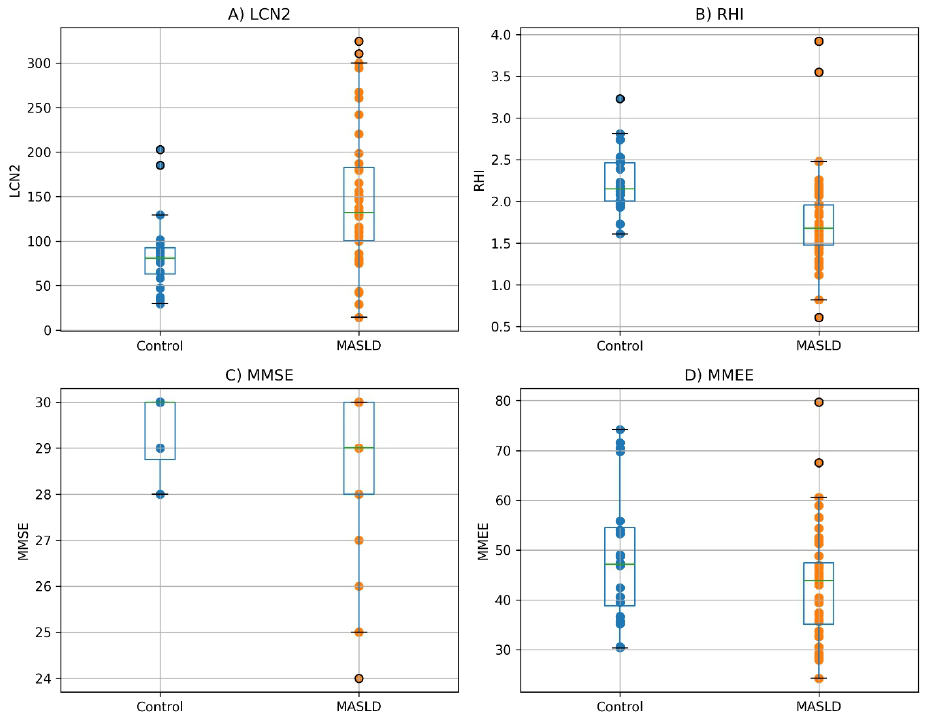
Figure 1. Distribution of LCN2, RHI, MMSE, and MMEE values in MASLD patients and controls. Box plots represent median and interquartile range, with individual data points overlaid (LCN2: lipocalin-2; RHI: reactive hyperemia index; MMSE: mini mental state examination; MMEE: myocardial mechano-energetic efficiency).
In addition, selected biochemical parameters were evaluated for descriptive purposes. Patients with MASLD showed higher ALT levels compared to controls (52.4 ± 18.7 vs. 24.8 ± 8.9 U/L). Similarly, total cholesterol and fasting glucose were higher in the MASLD group (212.6 ± 36.4 vs. 186.9 ± 28.7 mg/dL and 108.7 ± 14.8 vs. 89.6 ± 8.7 mg/dL, respectively), whereas HDL cholesterol levels were lower (41.3 ± 9.2 vs. 57.8 ± 11.4 mg/dL). These variables were not included in the predefined statistical analyses and are therefore reported as descriptive data only.
We also found that patients with MASLD were more commonly affected by hypertension (60% vs. 15%; p < 0.0005), dyslipidaemia (57.5% vs. 25%; p = 0.006), and diabetes mellitus (57.5% vs. 10%; p < 0.0005) than the controls were. With respect to drug therapies, MASLD patients were more likely to be treated with angiotensin-converting enzyme (ACE) inhibitors/angiotensin II receptor blockers (37.5% vs. 15%; p = 0.041), statins (35% vs. 14%; p = 0.039), beta-blockers (30% vs. 0%; p < 0.0005), calcium antagonists (15% vs. 0%; p = 0.026), sodium-glucose transporter II (SGLT2) inhibitors (12.5% vs. 0%; p = 0.021), metformin (50% vs. 10%; p < 0.0005) and insulin (12% vs. 0%; p = 0.021) than the control individuals were; no patients were taking drugs of the glucagon-like peptide 1-receptor agonist (GLP1-RA) class (Table 2).
Table 2. Comparison of comorbidities and drug therapy percentages between MASLD patients (N = 40) and control individuals (N = 40).
| Comorbidities and drug therapies | Patients, % | Controls, % | P-Value | ||||||||||||||||||||||||||||||||||||||||||||||||||||||||||||||||||||||||||||||||||||||||||||||||
| Hypertension | 60 | 15 | < 0.0005 | ||||||||||||||||||||||||||||||||||||||||||||||||||||||||||||||||||||||||||||||||||||||||||||||||
| Dyslipidaemia | 57.5 | 25 | 0.006 | ||||||||||||||||||||||||||||||||||||||||||||||||||||||||||||||||||||||||||||||||||||||||||||||||
| Diabetes mellitus | 57.5 | 10 | < 0.0005 | ||||||||||||||||||||||||||||||||||||||||||||||||||||||||||||||||||||||||||||||||||||||||||||||||
| ACE-I/ARB-II | 37.5 | 15 | 0.041 | ||||||||||||||||||||||||||||||||||||||||||||||||||||||||||||||||||||||||||||||||||||||||||||||||
| Statins | 35 | 14 | 0.039 | ||||||||||||||||||||||||||||||||||||||||||||||||||||||||||||||||||||||||||||||||||||||||||||||||
| Beta-blockers | 30 | 0 | < 0.0005 | ||||||||||||||||||||||||||||||||||||||||||||||||||||||||||||||||||||||||||||||||||||||||||||||||
| Calcium antagonists | 15 | 0 | 0.026 | ||||||||||||||||||||||||||||||||||||||||||||||||||||||||||||||||||||||||||||||||||||||||||||||||
| SGLT2-I | 12.5 | 0 | 0.021 | ||||||||||||||||||||||||||||||||||||||||||||||||||||||||||||||||||||||||||||||||||||||||||||||||
| Metformin | 50 | 10 | < 0.0005 | ||||||||||||||||||||||||||||||||||||||||||||||||||||||||||||||||||||||||||||||||||||||||||||||||
| Insulin | 12 | 0 | 0.021 | ||||||||||||||||||||||||||||||||||||||||||||||||||||||||||||||||||||||||||||||||||||||||||||||||
| ACE-I, angiotensin-converting enzyme inhibitor; ARB-II, angiotensin II receptor blocker; SGLT2-I, sodium-glucose transporter 2 inhibitor. | |||||||||||||||||||||||||||||||||||||||||||||||||||||||||||||||||||||||||||||||||||||||||||||||||||
Finally, we found no statistically significant differences between patients with MASLD and control individuals with respect to the IMT values of the carotid arteries according to ultrasound of the supra-aortic trunk.
The results of the multivariable regression analysis of the clinical, laboratory, and instrumental variables found to be significantly different between the groups in the univariable analysis indicated that, after adjusting for age, BMI and diabetes mellitus, the presence of MASLD was significantly positively associated with the LVPWT [OR=5.91; 95% CI: 1.43-24.5; p=0.014] and LCN2 [OR= 1.02 95% CI: 1.01-1.03, p=0.001] and significantly negatively associated with the RHI [OR= 0.18; 95% CI: 0.05-0.65, p=0.009] and MMSE score [OR=0.59; 95% CI: 0.36-0.98, p=0.048] (Tables 3–5).
Table 3. Results of multivariable logistic regression analysis for the relationships between significant clinical and laboratory variables according to univariable analysis (independent variables) and patient group (dependent variable).
| Variables | Adj. OR (95% CI) | P-value | |||||||||||||||||||||||||||||||||||||||||||||||||||||||||||||||||||||||||||||||||||||||||||||||||
| HR | 1.11 (0.98–1.23) | 0.082 | |||||||||||||||||||||||||||||||||||||||||||||||||||||||||||||||||||||||||||||||||||||||||||||||||
| BMI | 3.62 (1.27–10.3) | 0.016 | |||||||||||||||||||||||||||||||||||||||||||||||||||||||||||||||||||||||||||||||||||||||||||||||||
| BSA | 0.15 (0.01–10.3) | 0.382 | |||||||||||||||||||||||||||||||||||||||||||||||||||||||||||||||||||||||||||||||||||||||||||||||||
| HbA1c | 1.35 (0.32–5.66) | 0.682 | |||||||||||||||||||||||||||||||||||||||||||||||||||||||||||||||||||||||||||||||||||||||||||||||||
| MMSE | 0.59 (0.36–0.98) | 0.048 | |||||||||||||||||||||||||||||||||||||||||||||||||||||||||||||||||||||||||||||||||||||||||||||||||
| LCN2 | 1.02 (1.01–1.03) | 0.001 | |||||||||||||||||||||||||||||||||||||||||||||||||||||||||||||||||||||||||||||||||||||||||||||||||
| RHI | 0.18 (0.05–0.65) | 0.009 | |||||||||||||||||||||||||||||||||||||||||||||||||||||||||||||||||||||||||||||||||||||||||||||||||
| Diabetes mellitus | 10.8 (2.7–42.9) | 0.001 | |||||||||||||||||||||||||||||||||||||||||||||||||||||||||||||||||||||||||||||||||||||||||||||||||
| Age | 0.94 (0.88–1.01) | 0.063 | |||||||||||||||||||||||||||||||||||||||||||||||||||||||||||||||||||||||||||||||||||||||||||||||||
| Adj. Ors, adjusted odds ratios; BSA, body surface area; BMI, body mass index; CI, confidence interval; HbA1c, glycated hemoglobin; HR, heart rate; LCN2, lipocalin-2; RHI, reactive hyperemia index; MMSE, mini-mental state examination. | |||||||||||||||||||||||||||||||||||||||||||||||||||||||||||||||||||||||||||||||||||||||||||||||||||
| Reference: control group. | |||||||||||||||||||||||||||||||||||||||||||||||||||||||||||||||||||||||||||||||||||||||||||||||||||
| Data were adjusted for age, BMI and diabetes mellitus. | |||||||||||||||||||||||||||||||||||||||||||||||||||||||||||||||||||||||||||||||||||||||||||||||||||
Table 4. Results of multivariable logistic regression analysis for the relationships between significant echocardiographic variables according to univariable analysis (independent variables) and patient group (dependent variable).
| Variables | Adj. OR (95% CI) | P-value | |||||||||||||||||||||||||||||||||||||||||||||||||||||||||||||||||||||||||||||||||||||||||||||||||
| EF (%) | 0.94 (0.83–1.06) | 0.308 | |||||||||||||||||||||||||||||||||||||||||||||||||||||||||||||||||||||||||||||||||||||||||||||||||
| LAV | 0.96 (0.90–1.05) | 0.595 | |||||||||||||||||||||||||||||||||||||||||||||||||||||||||||||||||||||||||||||||||||||||||||||||||
| IVST | 0.67 (0.28–1.58) | 0.359 | |||||||||||||||||||||||||||||||||||||||||||||||||||||||||||||||||||||||||||||||||||||||||||||||||
| LVPWT | 5.91 (1.43–24.5) | 0.014 | |||||||||||||||||||||||||||||||||||||||||||||||||||||||||||||||||||||||||||||||||||||||||||||||||
| E/A | 0.15 (0.02–1.31) | 0.086 | |||||||||||||||||||||||||||||||||||||||||||||||||||||||||||||||||||||||||||||||||||||||||||||||||
| MMEE | 0.88 (0.87–1.00) | 0.065 | |||||||||||||||||||||||||||||||||||||||||||||||||||||||||||||||||||||||||||||||||||||||||||||||||
| BMI | 6.75 (1.57–29.0) | 0.010 | |||||||||||||||||||||||||||||||||||||||||||||||||||||||||||||||||||||||||||||||||||||||||||||||||
| Age | 0.97 (0.92–1.01) | 0.171 | |||||||||||||||||||||||||||||||||||||||||||||||||||||||||||||||||||||||||||||||||||||||||||||||||
| Diabetes mellitus | 1.61 (1.23–2.11) | 0.001 | |||||||||||||||||||||||||||||||||||||||||||||||||||||||||||||||||||||||||||||||||||||||||||||||||
| 95% CI, 95% confidence interval; Adj. Ors, adjusted odds ratios; E/A, e wave/a wave ratio; EF, ejection fraction; IVST, interventricular septal thickness; LAV, left atrial volume; LVPWT, left ventricular posterior wall thickness; MMEE, myocardial mechano-energetic efficiency. | |||||||||||||||||||||||||||||||||||||||||||||||||||||||||||||||||||||||||||||||||||||||||||||||||||
| Reference: control group. | |||||||||||||||||||||||||||||||||||||||||||||||||||||||||||||||||||||||||||||||||||||||||||||||||||
| Data were adjusted for age, BMI and diabetes mellitus. | |||||||||||||||||||||||||||||||||||||||||||||||||||||||||||||||||||||||||||||||||||||||||||||||||||
Table 5. Results of multivariable logistic regression analysis for the relationships between significant comorbidities and drug therapy variables according to univariable analysis (independent variables) and patient group (dependent variable).
| Variables | Adj. OR (95% CI) | P-value | |||||||||||||||||||||||||||||||||||||||||||||||||||||||||||||||||||||||||||||||||||||||||||||||||
| Hypertension | 4.65 (0.40–54.2) | 0.219 | |||||||||||||||||||||||||||||||||||||||||||||||||||||||||||||||||||||||||||||||||||||||||||||||||
| Dyslipidaemia | 0.94 (0.10–8.60) | 0.958 | |||||||||||||||||||||||||||||||||||||||||||||||||||||||||||||||||||||||||||||||||||||||||||||||||
| ACE-I/ARB II | 0.55 (0.07–4.10) | 0.561 | |||||||||||||||||||||||||||||||||||||||||||||||||||||||||||||||||||||||||||||||||||||||||||||||||
| Statins | 6.7 (0.75–59.8) | 0.088 | |||||||||||||||||||||||||||||||||||||||||||||||||||||||||||||||||||||||||||||||||||||||||||||||||
| Metformin | 2.70 (0.27–26.2) | 0.392 | |||||||||||||||||||||||||||||||||||||||||||||||||||||||||||||||||||||||||||||||||||||||||||||||||
| Age | 0.93 (0.86–1.01) | 0.059 | |||||||||||||||||||||||||||||||||||||||||||||||||||||||||||||||||||||||||||||||||||||||||||||||||
| BMI | 1.85 (1.34–2.56) | < 0.0005 | |||||||||||||||||||||||||||||||||||||||||||||||||||||||||||||||||||||||||||||||||||||||||||||||||
| Diabetes mellitus | 14.3 (1.42–52.3) | 0.017 | |||||||||||||||||||||||||||||||||||||||||||||||||||||||||||||||||||||||||||||||||||||||||||||||||
| 95% CI, 95% confidence interval; ACE-I, angiotensin-converting enzyme inhibitor; ARB II, angiotensin ii receptor blocker. | |||||||||||||||||||||||||||||||||||||||||||||||||||||||||||||||||||||||||||||||||||||||||||||||||||
| Reference: control group. | |||||||||||||||||||||||||||||||||||||||||||||||||||||||||||||||||||||||||||||||||||||||||||||||||||
| Data were adjusted for age, BMI and diabetes mellitus. | |||||||||||||||||||||||||||||||||||||||||||||||||||||||||||||||||||||||||||||||||||||||||||||||||||
Additional analyses were performed to further explore the relationships between LCN2 and vascular, cardiac, and cognitive parameters. Partial correlation analyses adjusted for age, BMI, and diabetes mellitus did not confirm statistically significant associations between LCN2 and RHI (r = -0.06, p = 0.73) or MMSE (r = 0.03, p = 0.86), while the association with MMEE showed a non-significant positive trend (r = 0.26, p = 0.12).
We also performed ROC curve analysis to explore the ability of RHI to predict MASLD; the results yielded an AUC of 0.826 (95% CI: 0.72–0.90; p<0.0005) at an optimal cut-off value of 1.87 (sensitivity=72.5%, specificity=90%), suggesting that the RHI can serve as a marker of endothelial dysfunction and thus as an indirect indicator of cardiovascular risk in patients with non-alcoholic fatty liver disease (Figure 2).
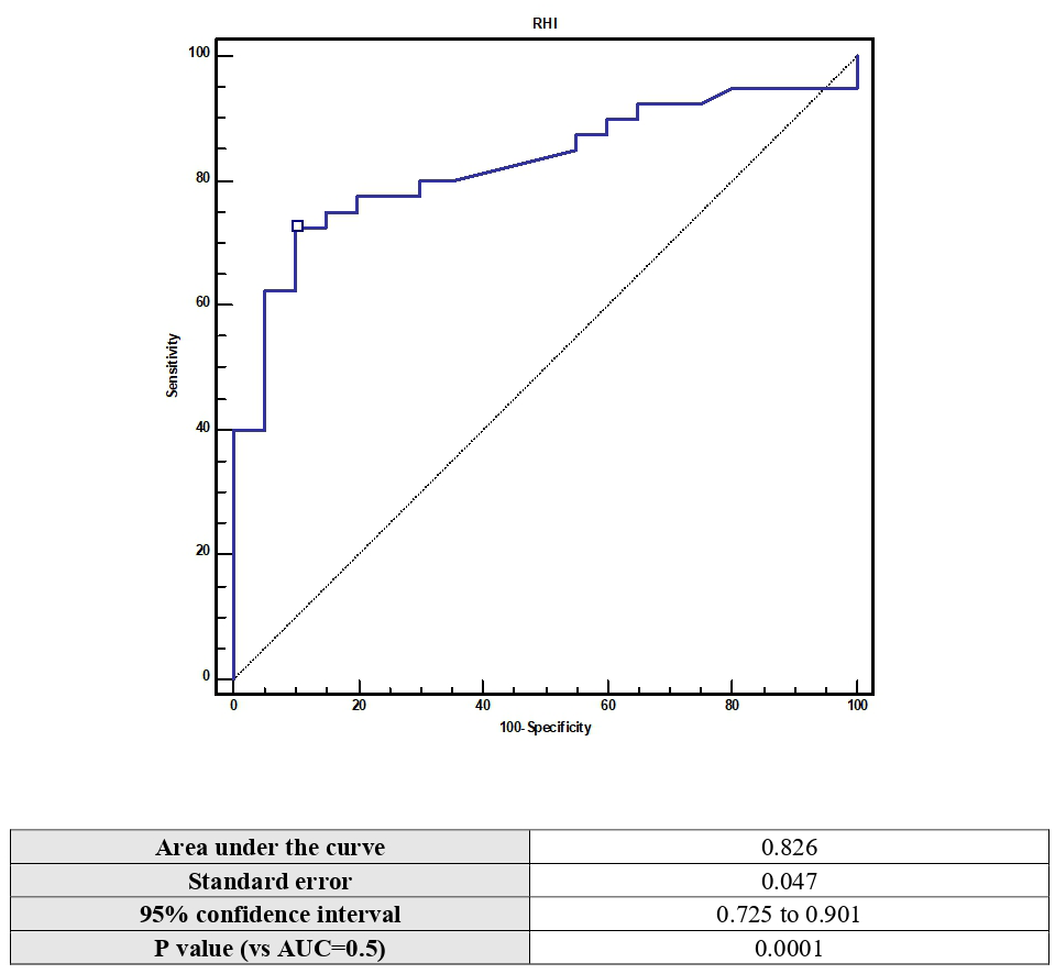
Figure 2. Receiver operating characteristic (ROC) curve and area under the curve (AUC) of the RHI in predicting NAFLD.
Discussion
In our study we found that, compared with controls, patients with MASLD had higher serum levels of LCN2, lower MMSE scores, and lower RHI values. We initially assumed an association between markers of adipo-neuroinflammation and hepatic steatosis (e.g., the LCN2 level), and surrogate indicators of cardiovascular risk (such as endothelial dysfunction as reflected in the RHI), indicators of cognitive impairment (such as the MMSE score), and indicators of myocardial performance (according to MMEE). Some previous studies evaluated only the arterial stiffness [25] or the cognitive performance of MASLD patients [26]; in a previous study comparing patients with MASLD with controls without hepatic steatosis, our group demonstrated that in the MASLD group, there was a negative correlation between indicators of cognitive impairment, such as the MMSE score, and histological markers of liver damage [14]. However, to date, no other clinical trials have attempted to assess the relationships among markers of neuroinflammation, cognitive impairment, and endothelial dysfunction.
Endothelial dysfunction is considered the starting point of vascular disease; through molecular mechanisms including vasoconstriction induced by the reduced availability of nitric oxide (NO) and inflammation, endothelial dysfunction leads to the onset of atherothrombotic events that drastically increase cardiovascular risk [27]. The RHI, obtained with the plethysmographic RH-PAT method, indirectly provides information on the degree of vasodilation after an induced hypoxia trigger; thus, the low RHI values found in patients with MASLD could indicate increased cardiovascular risk due to worse endothelial cell dysfunction.
Additionally, in MASLD patients, as reported above, we detected elevated serum levels of LCN2, which, on the basis of the available evidence in the literature, is a biohumoural marker of adipo-neuroinflammation. Several circulating inflammatory markers, such as C-reactive protein, interleukin-6, tumor necrosis factor-α, and adipokines, are recognised indicators of systemic inflammation in metabolic disorders. However, LCN2 was selected due to its ability to integrate metabolic, innate immune, vascular, and neuroinflammatory pathways. Consequently, LCN2 appears particularly appropriate for investigating the association between MASLD, endothelial dysfunction, and cognitive impairment within a unified pathophysiological framework.
In some mouse models of dysmetabolic-based inflammation (type 2 diabetes mellitus or nonalcoholic steatohepatitis), this protein promotes an inflammatory microenvironment through the recruitment of inflammatory cells (neutrophils) and the production of proinflammatory cytokines. It follows that LCN2 may play a role as a mediator of vascular damage in patients with MASLD, confirming a previously hypothesized theory proposing the presence of an adipose-liver-vascular axis linked to metabolic syndrome.
Other previous experimental evidences, instead, indicate that hepatocyte-specific overexpression of LCN2 protects against diet-induced steatosis in murine models [28]. This observation initially appears to contrast with the present findings. Several factors may explain this discrepancy. First, experimental models utilise controlled, cell-specific overexpression, whereas circulating LCN2 levels in humans represent the combined contributions of multiple tissues, including immune cells, adipose tissue, and vascular endothelium, within the context of systemic inflammation. Second, LCN2 may have compartment-specific and context-dependent effects, potentially providing protection within hepatocytes while serving as a marker or mediator of systemic inflammatory activation. Third, elevated circulating LCN2 in clinical settings likely reflects a compensatory or reactive response to chronic metabolic and inflammatory stress, rather than a primary protective mechanism. Finally, the function of LCN2 may differ across disease stages, exhibiting adaptive effects in early or controlled conditions and associations with adverse outcomes in chronic dysmetabolic states. Collectively, these considerations support interpreting LCN2 in this study primarily as a biomarker of systemic inflammatory burden, rather than as a direct causal driver of vascular and cognitive dysfunction.
LCN2 may serve as a mechanistic link between liver-centred metabolic inflammation and systemic vascular dysfunction. In dysmetabolic states, elevated circulating LCN2 likely indicates activation of innate immune pathways and enhanced cytokine signalling, which together sustain a pro-inflammatory environment that impairs endothelial homeostasis. Consequently, increased LCN2 levels are associated with reduced nitric oxide bioavailability, heightened oxidative stress, enhanced leukocyte recruitment, and a shift toward vasoconstrictive and pro-atherogenic conditions. These mechanisms align with the lower RHI values observed in the MASLD cohort.
In the group of patients with MASLD, we found a higher prevalence of risk factors falling within the framework of metabolic syndrome, which itself increases overall cardiovascular risk. The higher burden of cardiometabolic comorbidities observed in MASLD patients may have contributed to the differences in endothelial function, cardiac parameters, and LCN2 levels. Therefore, the observed associations likely reflect a complex interplay between liver disease and systemic metabolic dysfunction rather than a purely liver-specific effect.
Beyond the aggregation of traditional cardiometabolic risk factors, these findings support the interpretation of MASLD as a systemic inflammatory and metabolic disorder rather than an isolated liver condition. In the present study, MASLD can be interpreted as a clinical expression of systemic metabolic dysfunction, in which liver involvement coexists with cardiovascular and metabolic alterations. Within this context, hepatic steatosis leads to chronic low-grade inflammation, which promotes endothelial dysfunction and vascular impairment; insulin resistance alters glucose metabolism and heightens cardiovascular risk; adipose tissue dysfunction exacerbates metabolic imbalance; and vascular impairment further increases susceptibility to both cardiovascular and neurological injury. This systemic milieu establishes a biologically plausible connection between liver disease severity, endothelial dysfunction, and early cognitive changes, thereby linking hepatic inflammation and metabolic disturbances with both cardiovascular and neurological outcomes. Our finding of lower MMSE scores in patients with MASLD is consistent with that of previous studies that have shown an association among metabolic syndrome, arterial stiffness, and cognitive impairment. For example, a trial involving patients with nonalcoholic liver steatosis revealed lower brain volume through neuroimaging methods, suggesting that MASLD, considered a manifestation of metabolic syndrome, may play a role in brain ageing and consequently cognitive decline [29].
The use of the MMSE to indirectly assess cognitive performance allowed us to identify earlier cognitive deterioration in patients with MASLD than in controls, which could also reflect worse vascular health in the brain, wherein inflammation actively contributes to cerebral damage; in other words, the inflammatory microenvironment that is typical of MASLD and supposedly linked to LCN2 is also evident with premature brain ageing and faster cognitive impairment.
Experimental evidence indicates that circulating LCN2 interacts with the central nervous system; however, the mechanisms underlying its passage across the blood–brain barrier (BBB) remain undefined. LCN2 receptors, including 24p3R and megalin, are present on brain endothelial and neural cells, which supports their role in neurovascular signalling. Elevated circulating LCN2 levels correlate with BBB dysfunction and neuroinflammation, suggesting a liver–brain axis [29].
Additionally, LCN2 can be produced locally within the central nervous system by astrocytes, microglia, neurons, and endothelial cells in response to inflammatory stimuli. This suggests that the association between LCN2 and cognitive impairment may involve both peripheral and central sources, without necessarily requiring direct BBB translocation.
The association with cognitive performance can be understood within the same systemic framework. If LCN2 reflects or contributes to persistent low-grade inflammation, endothelial dysfunction, and microvascular injury, elevated levels may identify patients in whom metabolic liver disease is accompanied by a less favourable cerebral environment. Therefore, cognitive impairment in MASLD is unlikely to result from a single liver-specific mechanism. Instead, it likely arises from the combined effects of systemic inflammation, vascular dysfunction, and metabolic dysregulation, with LCN2 serving as a potential mediator or as an indicator of this adverse biological profile.
While these mechanisms support a potential pathophysiological role of LCN2, the extent to which this association is independent of the underlying metabolic profile remains uncertain. In the present study, additional analyses adjusting for age, BMI, and diabetes mellitus did not confirm independent associations between LCN2 and endothelial or cognitive parameters. This finding suggests that the observed relationships may be largely mediated by the underlying cardiometabolic profile rather than representing a direct and independent effect of LCN2. Within this framework, LCN2 should be interpreted primarily as a marker of systemic metabolic and inflammatory burden, reflecting the integrated impact of metabolic dysfunction on vascular and neurocognitive domains.
We developed multivariable logistic regression models incorporating variables found to be statistically significant in the invariable analysis to corroborate the associations with MASLD and confirmed a statistically significant association with low RHI levels, low MMSE scores and high serum LCN2 levels. These findings are in line with our previous results and suggest that patients with nonalcoholic hepatic steatosis could have a more pronounced endothelial dysfunction and early or subclinical cognitive differences than controls [14]; however, given the type of study and the small sample size, we cannot conclude that LCN2 is a mechanistic driver of vascular damage, we can only speculate on the possible role this mediator might play.
This consideration is clinically significant for interpreting the present data. Although the observed association between elevated serum LCN2, reduced RHI values, and lower MMSE scores aligns with a potential pathogenic role of this molecule, the cross-sectional study design precludes the determination of whether LCN2 is causally implicated in endothelial injury and cognitive decline or primarily reflects the severity of the underlying systemic inflammatory and metabolic burden in MASLD. Consequently, in this study, LCN2 should be regarded primarily as a biomarker indicative of a more severe extrahepatic phenotype. Its direct causal contribution to cardiovascular and neurological outcomes requires clarification through longitudinal and mechanistic investigations.
Given the dysmetabolic nature of MASLD, these data are in agreement with what has been previously reported in the medical literature, namely, the presence of an adiponeurovascular axis for nonalcoholic hepatic steatosis that is dependent on the proinflammatory activity of lipocalin 2 that arises in the context of metabolic dysregulation and influences vascular and cerebral health. Unsurprisingly, recent studies have proposed the use of the levels of LCN2 present in the cerebrospinal fluid as a marker of vascular dementia in differential diagnoses from other forms of neurodegenerative dementia, such as Alzheimer’s disease, as the evidences clearly indicates that this protein is produced under inflammatory and ischaemic conditions in the brain [30].
LCN2 also appears to play a role in the bowel in the regulation of the microbiota, where it exerts anti-inflammatory activity, but in all other parts of the body, evidence suggests that it has proinflammatory activity, in line with our data.
Although LCN2 represents a biologically plausible mediator linking metabolic inflammation, endothelial dysfunction, and neurocognitive alterations, the lack of evidence for a causal role in our study does not allow us to infer whether targeting LCN2 would translate into clinical benefit. At present, LCN2 should be primarily considered a marker of systemic inflammatory burden and disease severity, and its potential as a therapeutic target warrants further investigation in mechanistic and interventional studies.
Given these findings, we performed ROC curve analysis (Figure 1) and found that the endothelial dysfunction index RHI significantly discriminated patients with MASLD from controls: in patients with MASLD, this marker could be therefore an indicator of vascular damage, which is most likely due to the microenvironment of inflammation promoted by LCN2, which results in a shift in the balance between proconstrictive and prodilating factors - as is the case during homeostasis - toward the former in MASLD.
Furthermore, we found that patients with MASLD had higher heart rates and lower MMEE values than healthy controls did. These findings, particularly those close to the threshold of statistical significance, should be interpreted with caution given the number of comparisons performed. Several studies have shown that a reduced MMEE is associated with insulin resistance, obesity and type 2 diabetes mellitus and is considered an independent risk factor for cardiovascular events [16, 17]. Fiorentino et al. [19] demonstrated lower values of MMEE in patients with MASLD and that the relationship between MASLD and low MMEE was dependent on insulin resistance. Our MASLD patients had higher BMIs and HbA1c values and thus a greater prevalence of obesity and type 2 diabetes mellitus than the control patients did, which might explain their lower MMEE values. Moreover, patients with MASLD had some interesting echocardiographic findings compared with the control patients, including a thicker interventricular septum and posterior wall, a smaller EF and a reduced E/A ratio, all of which are indicative of early signs of hypertensive cardiopathy and diastolic dysfunction. In our study, patients with hepatic steatosis were more likely to be hypertensive and have diabetes mellitus than the controls were, possibly explaining why these patients present with echocardiographic signs of hypertensive heart disease and diastolic dysfunction, as widely demonstrated in the literature [31].
In conclusion, the results of our study show that MASLD can no longer be considered solely a liver disease without systemic impact, but rather should be thought of as a disease that affects all organs and systems of the body; within this systemic interpretation of MASLD, elevated LCN2 may facilitate the identification of patients with broader inflammatory, endothelial, and neurocognitive involvement. However, these results should not be overinterpreted as evidence of causality. Instead, the findings support the view that LCN2 represents a promising marker of disease complexity and extrahepatic risk, warranting further validation as both a stratification biomarker and a potential therapeutic target.
If, therefore, the vascular health in these patients is impaired, endothelial function would be damaged through the mechanisms described above, allowing us to understand the implications for overall cardio- and cerebrovascular risk. This suggests the need for targeted, patient-centred interventions that aims to modify their prognostic trajectory by acting on factors that can slow or reverse endothelial dysfunction, potentially reducing the incidence of future vascular events, but such interventions would need be evaluated in randomized controlled trials involving relatively large patient cohorts.
Interventions targeting the metabolic syndrome underlying MASLD, including lifestyle modification and treatments aimed at improving insulin resistance and cardiometabolic risk, may potentially affect endothelial function, systemic inflammation, and cognitive performance. However, our study was not designed to assess whether changes in MMSE, RHI, or LCN2 may occur in response to these interventions, and this question should be addressed in future longitudinal and interventional studies.
Limitations
Undoubtedly, our work has several limitations. First, the sample size was small, limiting the generalizability of the findings to the general population, to other ethnic groups, and to patients with more advanced liver disease. Some estimates showed wide confidence intervals, reflecting limited sample size and potential model instability. In addition, the presence of residual confounding related to cardiometabolic comorbidities and pharmacological treatments cannot be excluded. Although we performed multivariable analyses, the relatively small sample size limited the number of covariates that could be included without increasing the risk of model overfitting and unstable estimates. Consequently, it was not possible to fully adjust for all relevant cardiometabolic factors, and the observed associations should be interpreted with caution within the context of the overall metabolic profile. In addition, the lack of correction for multiple testing may have increased the risk of type I error. Second, the study design does not allow us to demonstrate the existence of a causal link among the increase in LCN2 level, endothelial dysfunction, and the presence of MASLD. In addition, since this is a single-centre study with a relatively geographically limited population, we cannot consider our findings to be valid for any population of patients with this disease, as there may be other factors in addition to those we examined that can influence disease severity, cardiovascular risk, and patient metabolic profiles. However, to our knowledge, we are the first to evaluate the associations among MASLD, endothelial dysfunction, cognitive impairment, and increased cardiovascular risk, all possibly related to increased serum levels of LCN2. Therefore, we set the stage for establishing new considerations with respect to the inflammatory microenvironment of patients with nonalcoholic liver disease, understanding the role of LCN2 in assessing cardiovascular risk in these patients, and potentially leveraging, if properly assessed and tested, this laboratory marker as a diagnostic tool in the early stages of the disease.
Author Contributions
G.P. and A.D.C.: conceptualization, study design, patient enrollment, data collection, and manuscript drafting; M.G.B.: vascular ultrasound assessments, data acquisition; A.C.: statistical analysis and data interpretation; D.C., G.M., V.D.C., M.D., L.A. and M.C.: data curation, laboratory analyses coordination, and manuscript editing; S.P., A.C., G.P., C.C., T.D.C., I.S., and R.P.: supervision, critical revision for important intellectual content, and validation of clinical methodology; D.D.R. and A.T.: oversight of cardiovascular assessments, interpretation of cardiometabolic findings, and manuscript refinement. All authors read and approved the final manuscript.
Conflicts of Interest
The authors declare that they have no conflicts of interest.
Ethical Statement and Consent
This cross-sectional study was conducted in accordance with the ethical principles of the Declaration of Helsinki and was approved by the Institutional Ethics Committee of Policlinico “Paolo Giaccone” (Palermo 1) (Approval No. “2023.04”, Date [19 Apr 2023]). Written informed consent was obtained from all participants prior to enrollment. All procedures were performed in accordance with relevant guidelines and regulations. Participant confidentiality was protected, and all data were anonymized before analysis.
Funding
No funding has been provided for this project.
References
- 1. Baffy G, Brunt EM, Caldwell SH. Hepatocellular carcinoma in non-alcoholic fatty liver disease: an emerging menace. J Hepatol. 2012; 56:1384–91. https://doi.org/10.1016/j.jhep.2011.10.027 [PubMed]
- 2. Wainwright P, Byrne CD. Bidirectional Relationships and Disconnects between NAFLD and Features of the Metabolic Syndrome. Int J Mol Sci. 2016; 17:367. https://doi.org/10.3390/ijms17030367 [PubMed]
- 3. Eslam M, Sanyal AJ, George J, and International Consensus Panel. MAFLD: A Consensus-Driven Proposed Nomenclature for Metabolic Associated Fatty Liver Disease. Gastroenterology. 2020; 158:1999–2014.e1. https://doi.org/10.1053/j.gastro.2019.11.312 [PubMed]
- 4. Chien MH, Ying TH, Yang SF, Yu JK, Hsu CW, Hsieh SC, Hsieh YH. Lipocalin-2 induces apoptosis in human hepatocellular carcinoma cells through activation of mitochondria pathways. Cell Biochem Biophys. 2012; 64:177–86. https://doi.org/10.1007/s12013-012-9370-1 [PubMed]
- 5. Asimakopoulou A, Borkham-Kamphorst E, Henning M, Yagmur E, Gassler N, Liedtke C, Berger T, Mak TW, Weiskirchen R. Lipocalin-2 (LCN2) regulates PLIN5 expression and intracellular lipid droplet formation in the liver. Biochim Biophys Acta. 2014; 1842:1513–24. https://doi.org/10.1016/j.bbalip.2014.07.017 [PubMed]
- 6. Chung IH, Chen CY, Lin YH, Chi HC, Huang YH, Tai PJ, Liao CJ, Tsai CY, Lin SL, Wu MH, Chen CY, Lin KH. Thyroid hormone-mediated regulation of lipocalin 2 through the Met/FAK pathway in liver cancer. Oncotarget. 2015; 6:15050–64. https://doi.org/10.18632/oncotarget.3670 [PubMed]
- 7. Krizanac M, Mass Sanchez PB, Weiskirchen R, Asimakopoulos A. A Scoping Review on Lipocalin-2 and Its Role in Non-Alcoholic Steatohepatitis and Hepatocellular Carcinoma. Int J Mol Sci. 2021; 22:2865. https://doi.org/10.3390/ijms22062865 [PubMed]
- 8. Lee S, Lee J, Kim S, Park JY, Lee WH, Mori K, Kim SH, Kim IK, Suk K. A dual role of lipocalin 2 in the apoptosis and deramification of activated microglia. J Immunol. 2007; 179:3231–41. https://doi.org/10.4049/jimmunol.179.5.3231 [PubMed]
- 9. Lee S, Kim JH, Kim JH, Seo JW, Han HS, Lee WH, Mori K, Nakao K, Barasch J, Suk K. Lipocalin-2 Is a chemokine inducer in the central nervous system: role of chemokine ligand 10 (CXCL10) in lipocalin-2-induced cell migration. J Biol Chem. 2011; 286:43855–70. https://doi.org/10.1074/jbc.M111.299248 [PubMed]
- 10. Tan Q, Zhang C, Rao X, Wan W, Lin W, Huang S, Ying J, Lin Y, Hua F. The interaction of lipocalin-2 and astrocytes in neuroinflammation: mechanisms and therapeutic application. Front Immunol. 2024; 15:1358719. https://doi.org/10.3389/fimmu.2024.1358719 [PubMed]
- 11. Dong M, Xi G, Keep RF, Hua Y. Role of iron in brain lipocalin 2 upregulation after intracerebral hemorrhage in rats. Brain Res. 2013; 1505:86–92. https://doi.org/10.1016/j.brainres.2013.02.008 [PubMed]
- 12. Naudé PJ, Nyakas C, Eiden LE, Ait-Ali D, van der Heide R, Engelborghs S, Luiten PG, De Deyn PP, den Boer JA, Eisel UL. Lipocalin 2: novel component of proinflammatory signaling in Alzheimer’s disease. FASEB J. 2012; 26:2811–23. https://doi.org/10.1096/fj.11-202457 [PubMed]
- 13. Abcouwer SF, Lin CM, Shanmugam S, Muthusamy A, Barber AJ, Antonetti DA. Minocycline prevents retinal inflammation and vascular permeability following ischemia-reperfusion injury. J Neuroinflammation. 2013; 10:149. https://doi.org/10.1186/1742-2094-10-149 [PubMed]
- 14. Tuttolomondo A, Petta S, Casuccio A, Maida C, Corte VD, Daidone M, Di Raimondo D, Pecoraro R, Fonte R, Cirrincione A, Zafonte R, Cabibi D, Cammà C, et al. Reactive hyperemia index (RHI) and cognitive performance indexes are associated with histologic markers of liver disease in subjects with non-alcoholic fatty liver disease (NAFLD): a case control study. Cardiovasc Diabetol. 2018; 17:28. https://doi.org/10.1186/s12933-018-0670-7 [PubMed]
- 15. Petta S, Tuttolomondo A, Gagliardo C, Zafonte R, Brancatelli G, Cabibi D, Cammà C, Di Marco V, Galvano L, La Tona G, Licata A, Magliozzo F, Maida C, et al. The Presence of White Matter Lesions Is Associated With the Fibrosis Severity of Nonalcoholic Fatty Liver Disease. Medicine (Baltimore). 2016; 95:e3446. https://doi.org/10.1097/MD.0000000000003446 [PubMed]
- 16. de Simone G, Izzo R, Losi MA, Stabile E, Rozza F, Canciello G, Mancusi C, Trimarco V, De Luca N, Trimarco B. Depressed myocardial energetic efficiency is associated with increased cardiovascular risk in hypertensive left ventricular hypertrophy. J Hypertens. 2016; 34:1846–53. https://doi.org/10.1097/HJH.0000000000001007 [PubMed]
- 17. Scheuermann-Freestone M, Madsen PL, Manners D, Blamire AM, Buckingham RE, Styles P, Radda GK, Neubauer S, Clarke K. Abnormal cardiac and skeletal muscle energy metabolism in patients with type 2 diabetes. Circulation. 2003; 107:3040–6. https://doi.org/10.1161/01.CIR.0000072789.89096.10 [PubMed]
- 18. Mancusi C, de Simone G, Best LG, Wang W, Zhang Y, Roman MJ, Lee ET, Howard BV, Devereux RB. Myocardial mechano-energetic efficiency and insulin resistance in non-diabetic members of the Strong Heart Study cohort. Cardiovasc Diabetol. 2019; 18:56. https://doi.org/10.1186/s12933-019-0862-9 [PubMed]
- 19. Fiorentino TV, Miceli S, Succurro E, Sciacqua A, Andreozzi F, Sesti G. Nonalcoholic fatty liver disease is associated with a decreased myocardial mechano-energetic efficiency. J Intern Med. 2021; 289:221–31. https://doi.org/10.1111/joim.13155 [PubMed]
- 20. Touboul PJ, Hennerici MG, Meairs S, Adams H, Amarenco P, Bornstein N, Csiba L, Desvarieux M, Ebrahim S, Hernandez Hernandez R, Jaff M, Kownator S, Naqvi T, et al. Mannheim carotid intima-media thickness and plaque consensus (2004-2006-2011). An update on behalf of the advisory board of the 3rd, 4th and 5th watching the risk symposia, at the 13th, 15th and 20th European Stroke Conferences, Mannheim, Germany, 2004, Brussels, Belgium, 2006, and Hamburg, Germany, 2011. Cerebrovasc Dis. 2012; 34:290–6. https://doi.org/10.1159/000343145 [PubMed]
- 21. Simon A, Gariepy J, Chironi G, Megnien JL, Levenson J. Intima-media thickness: a new tool for diagnosis and treatment of cardiovascular risk. J Hypertens. 2002; 20:159–69. https://doi.org/10.1097/00004872-200202000-00001 [PubMed]
- 22. Mazumder PK, O’Neill BT, Roberts MW, Buchanan J, Yun UJ, Cooksey RC, Boudina S, Abel ED. Impaired cardiac efficiency and increased fatty acid oxidation in insulin-resistant ob/ob mouse hearts. Diabetes. 2004; 53:2366–74. https://doi.org/10.2337/diabetes.53.9.2366 [PubMed]
- 23. Folstein MF, Folstein SE, McHugh PR. “Mini-mental state”. A practical method for grading the cognitive state of patients for the clinician. J Psychiatr Res. 1975; 12:189–98. https://doi.org/10.1016/0022-3956(75)90026-6 [PubMed]
- 24. Tuttolomondo A, Casuccio A, Guercio G, Maida C, Del Cuore A, Di Raimondo D, Simonetta I, Di Bona D, Pecoraro R, Della Corte V, Gulotta E, Gulotta G, Pinto A. Arterial stiffness, endothelial and cognitive function in subjects with type 2 diabetes in accordance with absence or presence of diabetic foot syndrome. Cardiovasc Diabetol. 2017; 16:2. https://doi.org/10.1186/s12933-016-0483-5 [PubMed]
- 25. Sunbul M, Agirbasli M, Durmus E, Kivrak T, Akin H, Aydin Y, Ergelen R, Yilmaz Y. Arterial stiffness in patients with non-alcoholic fatty liver disease is related to fibrosis stage and epicardial adipose tissue thickness. Atherosclerosis. 2014; 237:490–3. https://doi.org/10.1016/j.atherosclerosis.2014.10.004 [PubMed]
- 26. Seo SW, Gottesman RF, Clark JM, Hernaez R, Chang Y, Kim C, Ha KH, Guallar E, Lazo M. Nonalcoholic fatty liver disease is associated with cognitive function in adults. Neurology. 2016; 86:1136–42. https://doi.org/10.1212/WNL.0000000000002498 [PubMed]
- 27. Wang X, He B. Endothelial dysfunction: molecular mechanisms and clinical implications. MedComm (2020). 2024; 5:e651. https://doi.org/10.1002/mco2.651 [PubMed]
- 28. Xu Y, Zhu Y, Jadhav K, Li Y, Sun H, Yin L, Kasumov T, Chen X, Zhang Y. Lipocalin-2 Protects Against Diet-Induced Nonalcoholic Fatty Liver Disease by Targeting Hepatocytes. Hepatol Commun. 2019; 3:763–75. https://doi.org/10.1002/hep4.1341 [PubMed]
- 29. Weinstein G, Zelber-Sagi S, Preis SR, Beiser AS, DeCarli C, Speliotes EK, Satizabal CL, Vasan RS, Seshadri S. Association of Nonalcoholic Fatty Liver Disease With Lower Brain Volume in Healthy Middle-aged Adults in the Framingham Study. JAMA Neurol. 2018; 75:97–104. https://doi.org/10.1001/jamaneurol.2017.3229 [PubMed]
- 30. Llorens F, Hermann P, Villar-Piqué A, Diaz-Lucena D, Nägga K, Hansson O, Santana I, Schmitz M, Schmidt C, Varges D, Goebel S, Dumurgier J, Zetterberg H, et al. Cerebrospinal fluid lipocalin 2 as a novel biomarker for the differential diagnosis of vascular dementia. Nat Commun. 2020; 11:619. https://doi.org/10.1038/s41467-020-14373-2 [PubMed]
- 31. Petta S, Argano C, Colomba D, Cammà C, Di Marco V, Cabibi D, Tuttolomondo A, Marchesini G, Pinto A, Licata G, Craxì A. Epicardial fat, cardiac geometry and cardiac function in patients with non-alcoholic fatty liver disease: association with the severity of liver disease. J Hepatol. 2015; 62:928–33. https://doi.org/10.1016/j.jhep.2014.11.030 [PubMed]