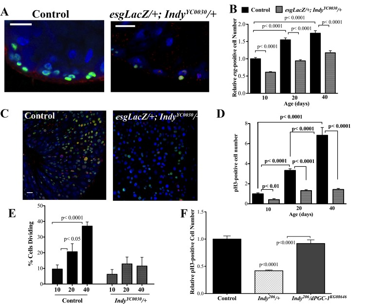
Figure 3.Indy mutations preserve ISC homeostasis. (A) Immunostaining for INDY (red) DAPI (blue nuclear) and β-galactosidase (green) in the midgut of female control (esgLacZ/+) and Indy mutant (esgLacZ;IndyYC0030/+) flies at 20 days at 40X. Scale bar represents 1μm. (B) Quantification shows reduced number of esg-positive cells in the esgLacZ; IndyYC0030/+ mutant female midgut throughout lifespan (p<0.0001, n>20). (C) Immunostaining for β-galactosidase activity (green), nuclear (blue) and pH3-positive cells (red) in control (esgLacZ/+) and the Indy mutant (esgLacZ/+;IndyYC0030/+) midgut tissue at 40 days. β-gal- positive cells represent ISC/EB populations and pH3-positive cells represent dividing cells. Scale bar represents 1μm. (D) Quantification of pH3-positive cells in the midgut of control (esgLacZ/+) and Indy (esgLacZ;IndyYC0030/+) mutant flies. There is increased cell division in female control midgut tissue throughout lifespan that is largely absent in esgLacZ; IndyYC0030/+ mutant females (p<0.01, p<0.0001, n>20). Error bars represent SEM. (E) Quantification of dividing cells in the midgut determined by the presence of pH3-positive immunostaining. There is increased cell division in female control midgut tissue throughout lifespan that is largely absent in esgLacZ; IndyYC0030/+ mutant females (p<0.05, p<0.0001, n>20). Error bars represent SEM. See Figures S4, S5 and Table S2. (F) Quantification of pH3- postive cells in the midgut of yw control, Indy206/+ mutant and Indy206/+/dPGC-1KG08646 female flies at 40 days. There are reduced dividing cells in the midgut of Indy mutant flies compared to control and Indy206/dPGC-1KG08646 midgut (p<0.0001, n>15). Error bars represent SEM.