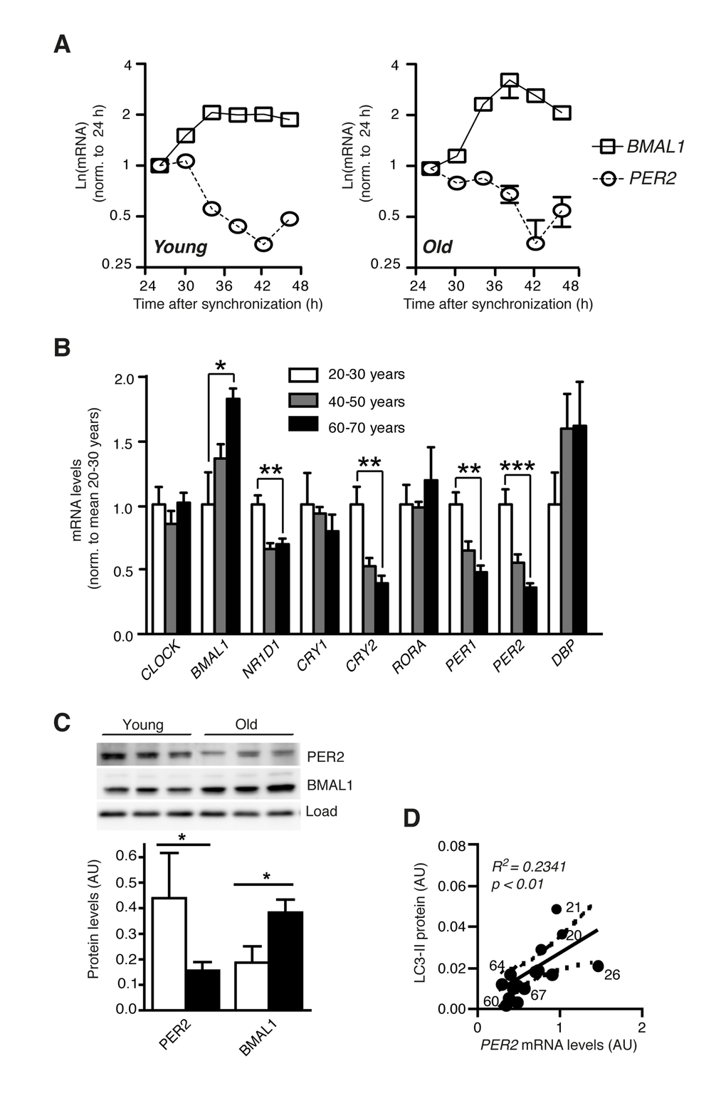
Figure 2.Clock gene expression is deregulated in aged primary human fibroblasts. (A) Circadian expression of Bmal1 mRNA (squares) and Per2 mRNA (circles) in primary human dermal fibroblasts from one young (age 21) and one old (age 67) donor of the cohort. (B) mRNA levels of core clock genes in cell lines from differently aged donors. Data obtained for age groups 20-30 years (white), 40-50 years (grey) and 60-70 years (black) are shown as mean ± SEM of five individual donors per age group. Data are normalised to the mean of age group 20-30 years. Asterisks indicate statistically significant differences between age groups 20-30 years and 60-70 years (unpaired t-test, two-tailed). (C) PER2 and BMAL1 protein levels in age groups 20-30 years (white) and 60-70 years (black). (D) Correlation of PER2 mRNA expression with LC3-II protein levels in the same cell lines. Solid and dashed lines indicate the linear regression curve and the 95% confidence band. Numbers next to data points show donor ages. Data are shown as mean values ± SEM, n=4.