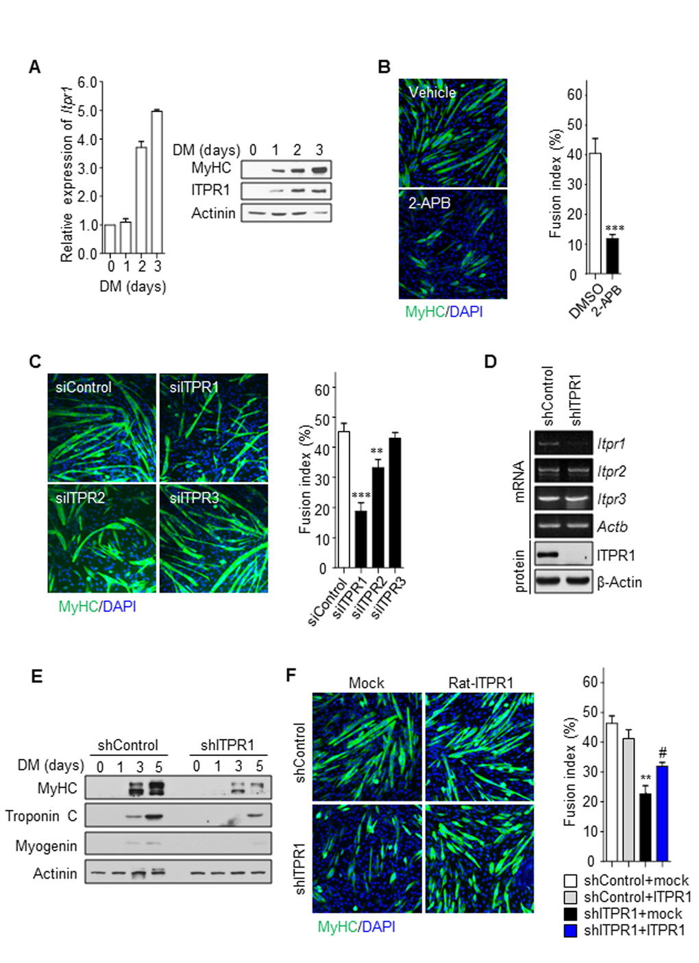
Figure 2.Inhibition of ITPR1 impairs myotube formation. (A) C2C12 myoblasts were induced to differentiate for 3 days and harvested at the indicated time-points. qRT-PCR and immunoblot analysis of mRNA and protein levels for ITPR1 in differentiating C2C12 cells. Actinin was used as the loading control. (B) C2C12 myoblasts were induced to differentiate for 5 days in the presence of 2-APB or vehicle and stained with MyHC (green) antibody and DAPI (blue). (C) C2C12 myoblasts were transfected with siRNA specific for each ITPR isoform or scrambled siRNA, induced to differentiate for 5 days, and stained with MyHC (green) antibody and DAPI (blue). (D) RT-PCR and immunoblot analyses of mRNA and protein levels of the three ITPR isoforms, respectively, in ITPR1-silenced C2C12 myoblasts. β-Actin was used as the loading control. (E) C2C12 myoblasts were transfected with siRNA against ITPR1 or scrambled siRNA. Cells were induced to differentiate for 5 days, harvested at the indicated time-points, and analyzed via immunoblotting with antibodies against MyHC, Troponin C and Myogenin. Actinin was used as the loading control. (F) Stable C2C12 cell lines expressing shRNA against ITPR1 or scrambled shRNA were transiently transfected with rat ITPR1 expression plasmids and induced to differentiate for 5 days, followed by staining with MyHC (green) antibody and DAPI (blue). The fusion index was calculated as the ratio of the number of multinucleated MyHC-positive myotubes to the number of total cells counted based on DAPI staining.