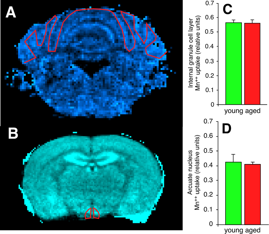
Figure 8.Synapses in the cerebellar internal granule cell layer of the aged C57BL/6 mouse and in the hypothalamic arcuate nucleus of the aged BALB mouse have functional deficits in excitatory amino acid neurotransmission. (A) Scaled difference MRI image of mouse cerebellum at approximately bregma -6.96 mm. Regions with higher Ca++ uptake are brighter on this colormap. The red line depicts the region-of-interest (ROI) drawn to include the internal granule cell layer. (B) Scaled difference MRI image of mouse hypothalamus at approximately bregma -1.46 mm. (C, D) Despite both regions demonstrating increased expression of regional-appropriate vesicular glutamate transporters, there is no evidence of increased post-synaptic Ca++ uptake in either the cerebellar internal granule cell layer (C) or the hypothalamic arcuate nucleus (D).