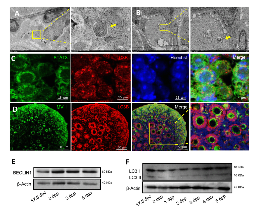
Figure 3.Morphological characteristics and expression of autophagic markers in mouse ovaries. (A) Observation of typical autophagosomes in 1 dpp mouse ovary by TEM. (B) Observation of typical autolysosome in 3 dpp mouse ovary by TEM. (C) Double IF staining for LC3B (red) and STAT3 (green) in 1 dpp mouse ovaries. (D) Double IF staining for LC3B (red) and MVH (green) in 3 dpp mouse ovaries. (E) WB detection for BECLIN1 in 17.5 dpc, 0 dpp, 3 dpp and 5 dpp mouse ovaries. (F) WB detection for BECLIN in 17.5 dpc, 0 - 5 dpp mouse ovaries.