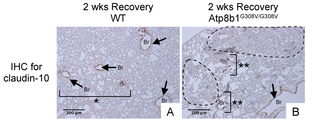
Figure 8.Atp8b1G308V/G308V mice exposed to hyperoxia and returned subsequently to normoxia for recovery show impaired bronchiolar regeneration. WT and Atp8b1G308V/G308V mice at 7-9 weeks of age were exposed to room air or 100% O2 for 48 hours, and then allowed to recover under normoxia for 12 days (n=3 for each). Representative photomicrographs of lung sections immunohistochemically labeled for claudin-10 are shown. Arrows denote relatively intact bronchiolar lumens. (A) WT lungs recovered from hyperoxia display normal regeneration of bronchioalveolar structures wherein organized arrangements of bronchioles surrounded by intact alveoli are noted (area designated by one asterisk). (B) Atp8b1G308V/G308V lungs recovered from hyperoxia display impaired regeneration of bronchioalveolar structures wherein incomplete bronchiolar structures (two asterisks) are juxtaposed to highly remodeled lesions (areas circled by dashed lines). Br: Bronchiolar lumen. Magnifications: (A and B) 200X. Data presented are representative of two independent experiments.