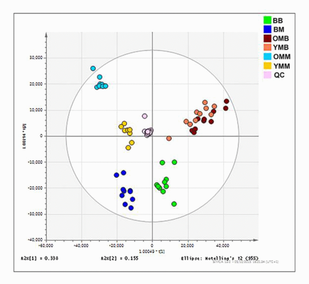
Figure 2.Mitochondrial lipid composition differs between the bat and mouse mitochondrial proteomes. Orthogonal partial least square-discriminant analysis (OPLS-DA) of lipids found in the brain and skeletal muscle mitochondria from the mouse and the bat. Separation across the x-axis is according to tissue type with the skeletal muscle mitochondrial samples congregating to the left quadrants and the brain mitochondrial samples to the right. Along the y-axis separation delineates mammalian species with the bat mitochondrial samples grouping at the lower quadrants and the mouse mitochondrial samples grouping at the upper quadrants. Bat brain (BB) mitochondrial samples (adult, n=10) are shown on the OPLS-DA by the green circles. Bat skeletal muscle (BM) mitochondria (adult, n=10) are indicated by the dark blue circles. Young mouse brain (YMB) mitochondria aged 4-11 weeks (n=10) and aged mouse brain mitochondria (OMB) aged 78 weeks (n=10) are denoted by orange and red circles respectively. Young mouse skeletal (YMM) muscle mitochondria aged 4-11 weeks (n=9) and aged mouse skeletal muscle mitochondria (OMM) aged 78 weeks (n=10) are indicated by the yellow and blue circles, respectively.