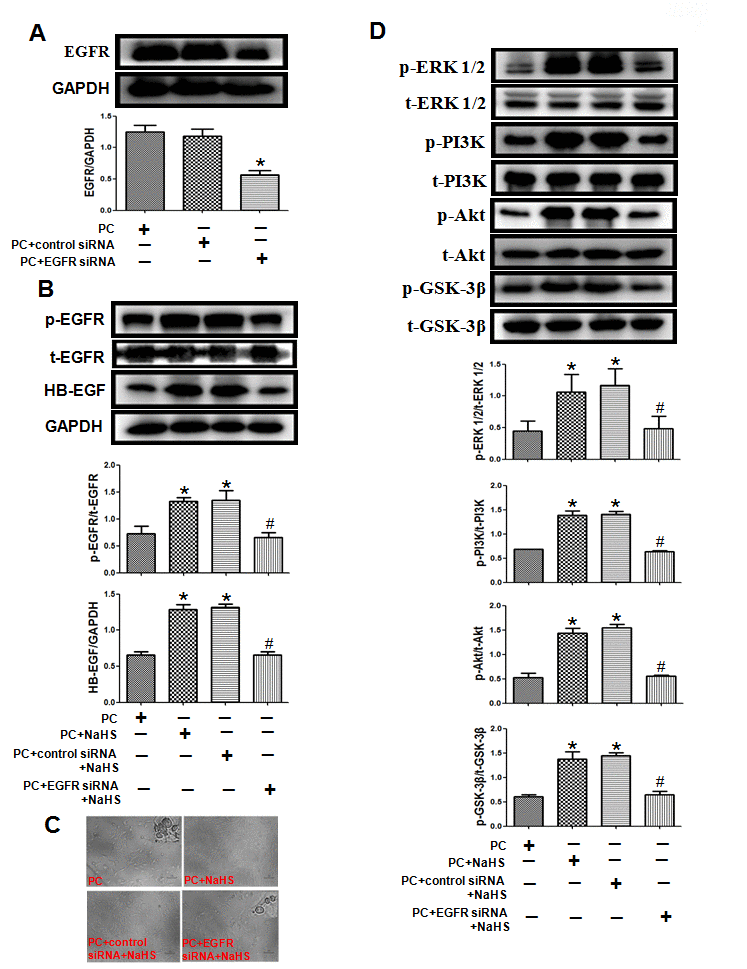
Figure 7.Knockdown of EGFR cancels the effect of exogenous H2S on cell damage and related signaling pathways in the aged H9C2 cells. (A) The knockdown of EGFR by EGFR-specific siRNA (EGFR siRNA) in the aged H9C2 cells. The cells were transfected with 50 nM EGFR siRNA or negative control siRNA (control siRNA) for 48 h during P(C) The data are the means ± S.E.M. of 3 determinations. * p<0.05 vs. PC + control siRNA group. (B) The knockdown of EGFR inhibited NaHS-increased expression of the HB-EGF and the activity of phosphorylated EGFR. The graphs represent the optical density of the bands of phosphorylated EGFR (p- EGFR) normalized to the expression of total EGFR (t-EGFR). The graphs represent the optical density of the bands of HB-EGF normalized to the expression of GAPDH signal. All data were from three independent experiments. * p<0.05 vs. PC group; # p<0.05 vs. PC + control siRNA + NaHS group. (C) The knockdown of EGFR cancelled NaHS-decreased cell damage. The cells were cultured in glass-bottom dishes and observed using a general inverted microscope (magnification ×100). (D) The knockdown of EGFR inhibited NaHS-up-regulated the ERK1/2 and PI3K-Akt-GSK-3β pathways. The phosphorylation of ERK1/2, PI3K, Akt and GSK-3β was detected using western blotting analysis. The graphs represent the optical density of the bands of phospho-ERK1/2 (p-ERK1/2), PI3K (p- PI3K), Akt (p-Akt) and GSK-3β (p-GSK-3β) normalized to the expression of total-ERK1/2 (t-ERK1/2) PI3K (t-PI3K), Akt (t-Akt) and GSK-3β (t-GSK-3β). All data were from three independent experiments. * p<0.05 vs. PC group; # p<0.05 vs. PC + control siRNA + NaHS group.