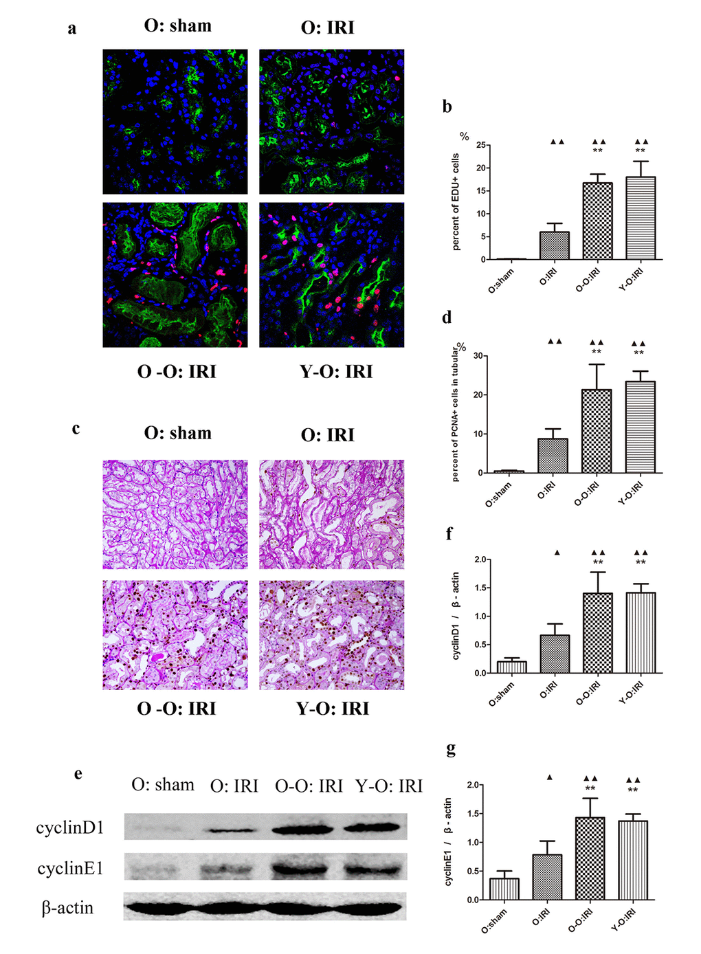
Figure 2.Exogenous biological renal support increased renal cell proliferation in old IRI mice. (A) Representative images of renal EdU-positive cells in independent groups (600× magnification; red, EDU; green, LTL; blue, DAPI). (B) The percentages of EdU-positive cells in the kidneys of the old mice at 72 hours after IRI. The mice in the O: IRI group displayed more EdU-positive cells than in O: sham group. The percentage of EdU-positive cells was higher in the O-O: IRI group and the Y-O: IRI group than in the O: IRI group. No significant difference was found between the O-O: IRI group and the Y-O: IRI group. (C) Representative images of renal PCNA-positive tubular cells in independent groups (400× magnification). (D) The percentages of PCNA-positive tubular cells in the kidneys of the old mice at 72 hours after IRI. The mice in the O: IRI group had more PCNA-positive tubular cells than the O: sham group. The percentages of PCNA-positive tubular cells were higher in the O-O: IRI group and the Y-O: IRI group than in the O: IRI group. No significant difference was found between the O-O: IRI group and the Y-O: IRI group. (E) The levels of cyclin D1 and cyclin E1 in kidney extracts of the old IRI mice as measured by western blotting. Gels were performed under the same experimental conditions. (F, G) Quantitative analyses of the band densities of cyclin D1 and cyclin E1 protein expression. Data are presented as means ± SDs. ▲P < 0.05, ▲▲P < 0.01 vs. O: sham; *P < 0.05, **P < 0.01 vs. O: IRI. SD, standard deviation.