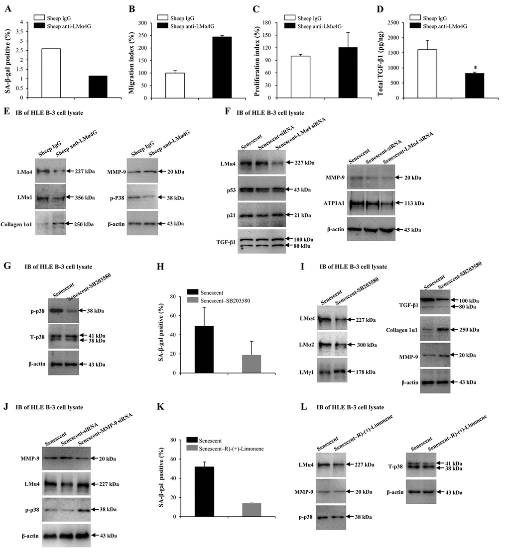
Figure 7.Interactions between laminin α4 (LMα4) and the activated p38 mitogen-activated protein kinase (p38 MAPK) signaling pathway in cell senescence. (A-E) Cells were treated with sheep anti-LMα4 globular domain antibodies (2 μg/ml) for 96 h, while cells treated with sheep IgG (2 μg/ml) were selected as the control group. (A) Percentage of SA-β-gal-positive cells. (B) Migratory abilities of HLE B-3 cells. (C) Cell viabilities of HLE B-3 cells measured by CCK-8 assay. (D) Total TGF-β1 in HLE B-3 cells detected by ELISA. (E) Immunoblot analysis of p-p38, collagen 1α1, MMP-9, and LMs in HLE B-3 cells. (F) HLE B-3 cells treated with 400 μM H2O2 for 96 h only, or in combination with indicated siRNA. Immunoblot analysis of LMα4, p21, p53, TGF-β1, MMP-9 and ATP1A1 in HLE B-3 cells. (G-I) HLE B-3 cells treated with H2O2 (400 μM) only for 96 h, or in combination with SB203580 (30 μM). (G) Immunoblot analysis of p-p38 and T-p38 (total p38) in HLE B-3 cells. (H) Percentage of SA-β-gal-positive cells. (I) Immunoblot analysis of TGF-β1, collagen 1α1, MMP-9 and LMs in HLE B-3 cells. (J) HLE B-3 cells were treated with H2O2 (400 μM) only for 96 h, or in combination with indicated siRNA. Immunoblot analysis of MMP-9, p-p38 and LMα4 in HLE B-3 cells. (K-L) HLE B-3 cells were treated with H2O2 (400 μM) only for 96 h, or in combination with R)-(+)-Limonene (1000 μM). (K) Percentage of SA-β-gal-positive cells. (L) Immunoblot analysis of p-p38, T-p38, MMP-9 and LMα4 in HLE B-3 cells. Data were shown as mean ± SD and were analyzed using the paired t-test. *, p<0.05.