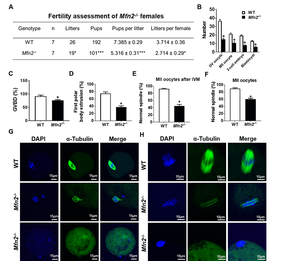
Figure 1.Subfertility, and impaired follicle, oocyte and embryo development in Mfn2-/- mice. (A) Fertility of female Mfn2-/- (oocyte-specific Mfn2 knockout, Mfn2fl/fl/Zp3-Cre, referred to as Mfn2-/-) and WT mice (8-week-old, n = 7 for each genotype) was assessed by mating with WT males of proven fertility (male/female; 1:2) for 12 weeks. Mfn2-/- mice had smaller litter size (pups per litter) and litters per female compared with WT females. (B) Number of GV oocytes, MII oocytes, 2-cell embryos and blastocysts in Mfn2-/- and WT mice. (C, D) Oocytes at GV stage were collected from PMSG-primed Mfn2-/- and WT mice and analyzed after 18 h of culture under in vitro maturation conditions. Percentages of GVBD and of first polar body extrusion in Mfn2-/- and WT oocytes are shown. (E, G) After 18 h of IVM, Mfn2-/- and WT MII oocytes were stained with α-tubulin and DAPI. Left column, DAPI (blue); middle column, anti-α-tubulin antibody (green); right column, merged images of DAPI and anti-α-tubulin staining. Percentages of normal spindle morphology in Mfn2-/- and WT MII oocytes after IVM are shown. (F, H) Mature (MII) oocytes were collected from the oviducts of superovulated 8-week-old Mfn2-/- and WT mice and stained with α-tubulin and DAPI. Percentages of normal spindle morphology in Mfn2-/- and WT MII oocytes are shown. Data presented as mean ± SEM. *p < 0.05, ***p < 0.0001, vs. WT using t-test.