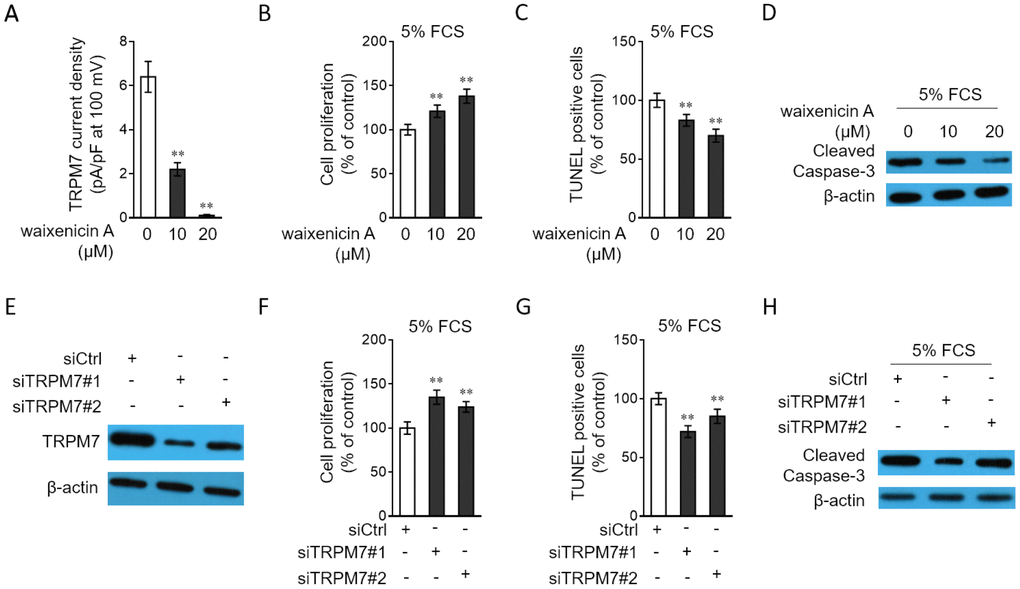
Figure 4.TRPM7 inhibition or knockdown promotes proliferation and apoptosis resistance in PASMCs. (A) PASMCs from rats were treated with vehicle control, 10 μM or 20 μM waixenicin A for 24 h. TRPM7 currentss were recorded using the whole-cell patch-clamp technology with ramp from -100 mV to 100 mV. TRPM7 currents density (pA/pF) with ramp at 100 mV is shown. Fifty cells were analyzed in each treatment. Data are mean ± SD. One-way ANOVA test. **, P < 0.01 compared to control. (B–D) PASMCs from rats were serum starved for 24 h, followed by incubation with medium containing 5% FCS for 24 h in the presence or absence of 10 μM or 20 μM waixenicin A. (B) Cell proliferation was determined by BrdU incorporation assay. (C) Cell apoptosis was detected by TUNEL staining. Results are expressed as a percentage relative to control. Data are mean ± SD. n = 3. One-way ANOVA test. **, P < 0.01 compared to control. (D) The protein expression of cleaved caspase-3 was determined by Western blot analysis. β-actin was used as a loading control. The representative images from 3 independent experiments are shown. (E) PASMCs from rats were transfected with siRNA control, siRNA TRPM7#1 or siRNA TRPM7#2. After 72 h of transfection, the protein level of TRPM7 was determined by Western blot analysis. β-actin was used as a loading control. The representative images from 3 independent experiments are shown. (F–H) PASMCs from rats were transfected with siRNA control, siRNA TRPM7#1 or siRNA TRPM7#2. After 24 h of transfection, cells were serum starved for 24 h, followed by incubation with medium containing 5% FCS for another 24 h. Cell proliferation (F), cell apoptosis (G) and the protein expression of cleaved caspase-3 (H) were determined and analyzed as in (B–D).