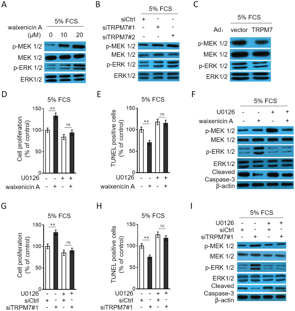
Figure 6.TRPM7 inhibition or knockdown promotes PASMCs proliferation and apoptosis resistance through MEK/ERK pathway. (A) PASMCs from rats were serum starved for 24 h, followed by incubation with medium containing 5% FCS for 24 h in the presence or absence of 10 μM or 20 μM waixenicin A. The protein expression of p-MEK 1/2, MEK 1/2, p-ERK 1/2 and ERK 1/2 was determined by Western blot analysis. (B) PASMCs from rats were transfected with siRNA control, siRNA TRPM7#1 or siRNA TRPM7#2. After 24 h of transfection, cells were serum starved for 24 h, followed by incubation with medium containing 5% FCS for another 24 h. The protein expression of p-MEK 1/2, MEK 1/2, p-ERK 1/2 and ERK 1/2 was determined by Western blot analysis. (C) PASMCs from rats were infected with Ad-Ctrl or Ad-TRPM7. After 24 h of infection, cells were serum starved for 24 h, followed by incubation with medium containing 5% FCS for another 24 h. The protein expression of p-MEK 1/2, MEK 1/2, p-ERK 1/2 and ERK 1/2 was determined by Western blot analysis. β-actin was used as a loading control. The representative images from 3 independent experiments are shown. (D–F) PASMCs from rats were serum starved for 24 h, followed by incubation with medium containing 5% FCS for 24 h in the presence or absence of 20 μM waixenicin A or 10 μM U0126. (D) Cell proliferation was determined by BrdU incorporation assay. (E) Cell apoptosis was detected by TUNEL staining. Results are expressed as a percentage relative to control. Data are mean ± SD. n = 3. Unpaired Student’s t-test. **, P < 0.01; NS, not significant, as compared to control. (F) The protein expression of targets as indicated was determined by Western blot analysis. β-actin was used as a loading control. The representative images from 3 independent experiments are shown. (G–I) PASMCs from rats were transfected with siRNA control, siRNA TRPM7#1. After 24 h of transfection, cells were serum starved for 24 h, followed by incubation with medium containing 5% FCS for another 24 h in the presence or absence of 10 μM U0126. Cell proliferation (G), cell apoptosis (H) and the protein expression of targets (I) was determined and analyzed as in (D–F).