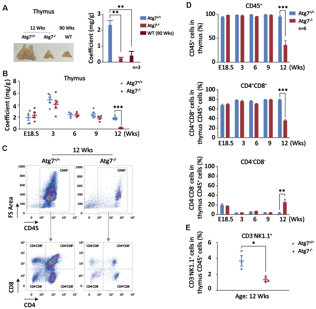
Figure 3.Autophagy defect causes synchronous thymic atrophy and T cell (CD4+CD8+) reduction after mouse development is completed. (A) Alteration of thymus in size and weight. Left, a representative picture of thymus; right, thymus coefficient of Atg7-deleted mice as compared with the same-age wild-type mice and the old wild-type mice. (B) Measurement of thymus coefficients in time points indicated in the entire lifespan of the Atg7-deleted mice (organ/body, mg/g). (C) Scheme for analysis of T cells by flow cytometry. Shown are representative flow images for quantification of total blood cell (CD45+) and T cells (CD4+,CD8+) in total thymus cells. (D) Statistical analysis of the percentages of T cell populations in total thymus blood cells in the entire lifespan of the Atg7-deleted mice. (E) Statistical analysis of the percentages of NK cell populations in total thymus blood cells in the Atg7-deleted mice at age of 12 weeks.