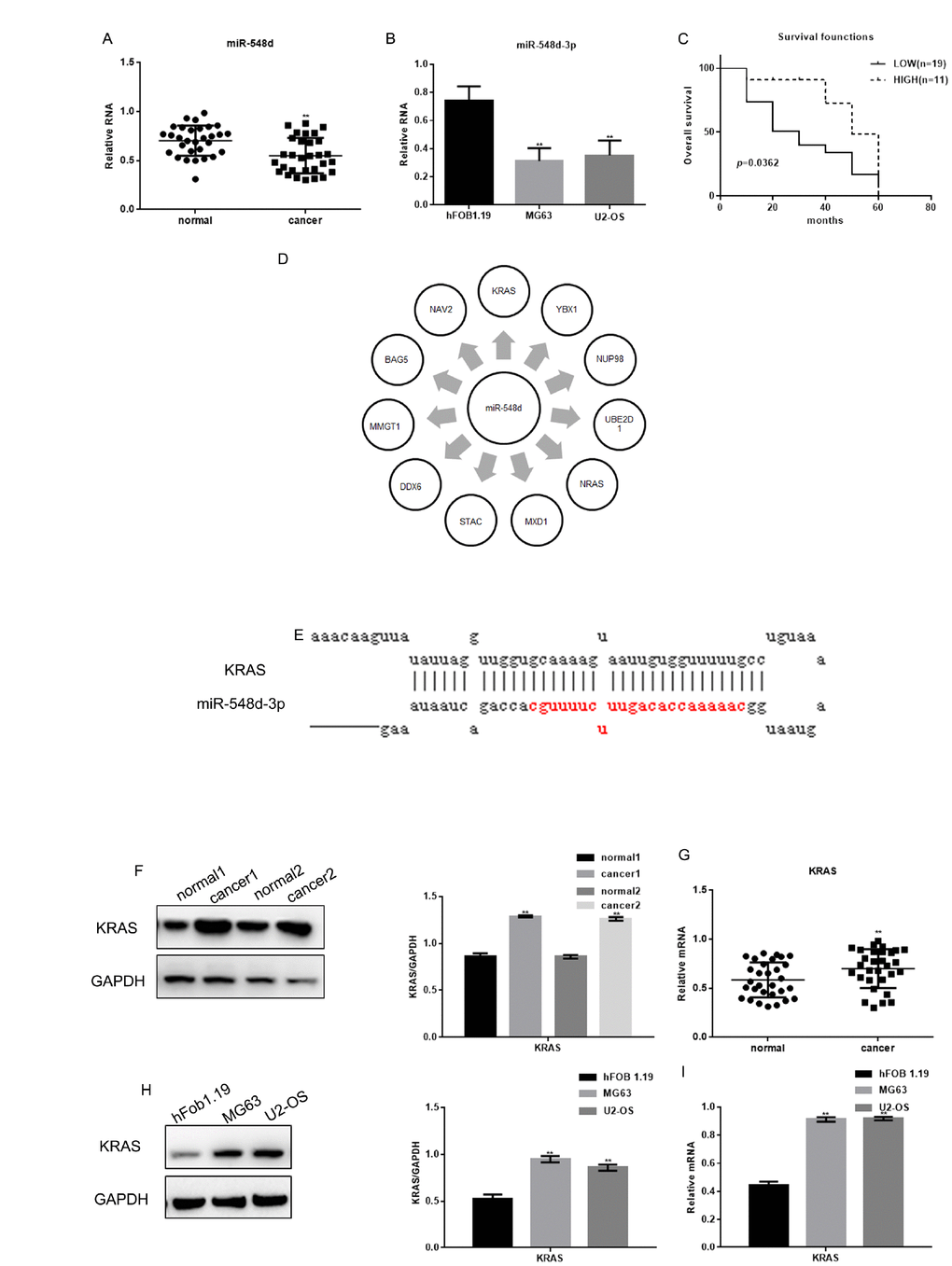
Figure 1.The expression of miR-548d-3p and KRAS in osteosarcoma. (A) The relative miR-548d-3p levels in 30 pairs of osteosarcoma tissues and adjacent normal tissues were detected by real-time PCR. Patients with lower miR-548d expression in their tumors than in mean of the normal. The results represent the mean±SD. **P < 0.05 vs. adjacent normal tissues. (B) Relative miR-548d-3p expression in cell lines (hFOB 1.19, MG63 and U2-OS). MG63 and U2-OS cells with lower miR-548d expression than hFOB 1.19 cells. The results represent the mean±SD of three independent experiments. **P < 0.05 vs. hFOB 1.19 cells. (C) The correlation between miR-548d-3p expression and patient survival. (D) Software analysis with miRDB revealed miR-548d-3p binding sites in a variety of genes. (E) The miRDB tool predicted that miR-548d-3p may bind to KRAS. (F) The relative KRAS levels in two pairs of osteosarcoma tissues and adjacent normal tissues were detected by Western blotting. The results represent the mean±SD. **P < 0.05 vs. adjacent normal tissues. (G) The relative KRAS levels in 30 pairs of osteosarcoma tissues and adjacent normal tissues were detected by Western blotting and real-time PCR. The results represent the mean±SD. **P < 0.05 vs. adjacent normal tissues. (H, I) The relative KRAS levels in cell lines (hFOB 1.19, MG63 and U2-OS) were detected by Western blotting and real-time PCR. The results represent the mean±SD of three independent experiments. **P < 0.05 vs. hFOB 1.19 cells.