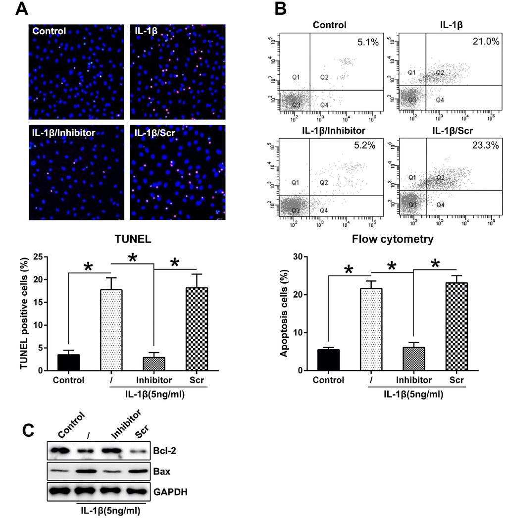
Figure 4.Downregulation of miR-27a expression repressed the apoptosis levels in IL-1β-triggered chondrocytes. (A) TUNEL staining was performed to detect the number of dead cells in case of IL-1β-treated chondrocytes that underwent transfection with the miR-27a inhibitor and miR-Scr. Magnification, ×100. The apoptotic rate based on positive TUNEL staining in each group is displayed in the lower right panel. (B) FC was performed to assess the number of dead cells that were transfected with the miR-27a inhibitor and miR-Scr. The upper right quadrant of every plot represents the early apoptotic cells. The apoptotic rate analysis of IL-1β-treated chondrocytes in each group is presented in the lower right panel. (C) WB was performed to detect the expression of the apoptotic markers, Bcl-2 and Bax, in IL-1β-treated chondrocytes. The results are described as the mean ± SD. *P < 0.05 vs. indicated group.