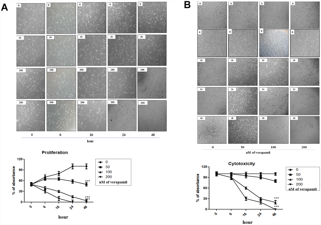
Figure 3.Cellular proliferation and viability by verapamil in hVFFs. Photographs and graphs show the changes of the proliferation (A) and viability (B) of hVFFs at various time points depending on the concentration of verapamil. Verapamil treatment reduced the proliferation and viability of hVFFs cells as dose- and time-dependent manner. Represented are light microscopic images of hVFFs for general morphology. One-way ANOVA test; ***p<0.001.