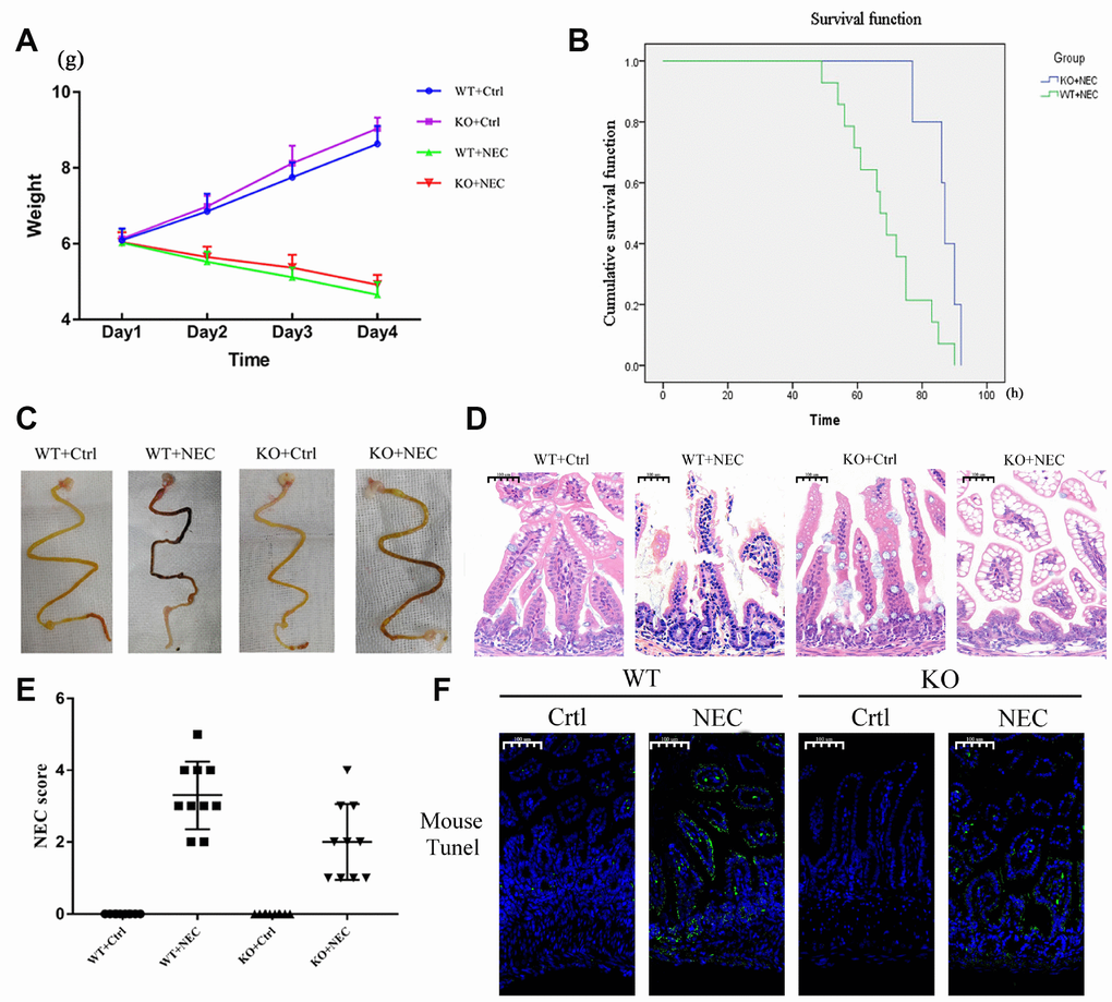
Figure 2.Targeted deletion of β-arrestin-2 impeded the development of NEC. (A) The body weights of mice decreased more slowly in the KO+NEC group (red) than in the WT+NEC group (green) (n=20). (B) The survival rate was much higher in the KO+NEC group (blue) than in the WT+NEC group (green) (n=20). (C) Representative images of gross morphology demonstrate that edema, congestion, necrosis and reddish-black coloring were more severe in the WT+NEC group than in the KO+NEC group. (D) Representative images display the more serious histological changes in the WT+NEC group, including the shedding of epithelial cells, necrosis of the entire villus and transmural necrosis. (E) The NEC score was lower in the KO+NEC group (2.00±1.05) than in the WT+NEC group (3.30±0.95) (P=0.01) (n=20). (F) A TUNEL assay revealed a much lower proportion of apoptotic cells in the KO+NEC group than in the WT+NEC group.