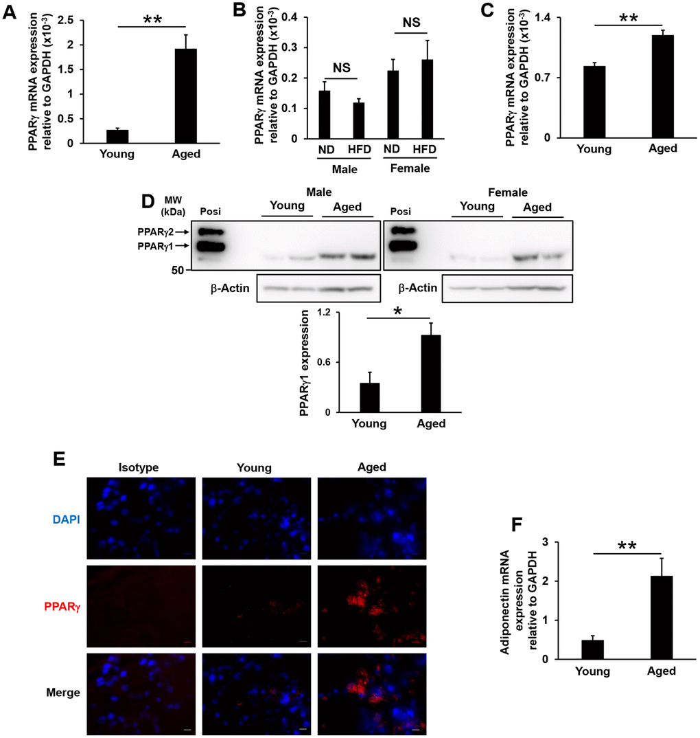
Figure 4.PPARγ expression in lacrimal glands and adiponectin mRNA expression in the white adipose tissue of aged and high-fat diet-fed mice. (A and B) PPARγ mRNA expression levels in the lacrimal glands of young and aged mice (N=7-8) (A), or of mice fed a normal diet (ND) or high-fat diet (HFD) for 8 weeks (N=4-5) (B). (C) PPARγ mRNA expression levels in the epithelial cells of the lacrimal glands of young and aged mice (N=4). (D) Detection of the PPARγ protein by Western blotting. Lysates prepared from the lacrimal glands of young and aged mice were immunoblotted with anti-AdipoR2 and anti-β-Actin antibodies. Left and right images show male (N=2) and female mice (N=2), respectively. The positive control (Posi) is a lysate prepared from the subcutaneous fat of young mice. The bar graph shows integrated signal intensities in AdipoR2 normalized to that of β-Actin (N=4). (E) PPARγ expression in the acinar cells of the lacrimal glands of young and aged mice as detected by immunofluorescence. Nuclei were stained with DAPI. Bars = 10 μm. (F) Adiponectin mRNA expression levels in the mesenteric white adipose tissues of young and aged mice (N=4-5). Values are presented as means ± SEM. NS, not significant. **p<0.01 (an unpaired Student’s t-test).