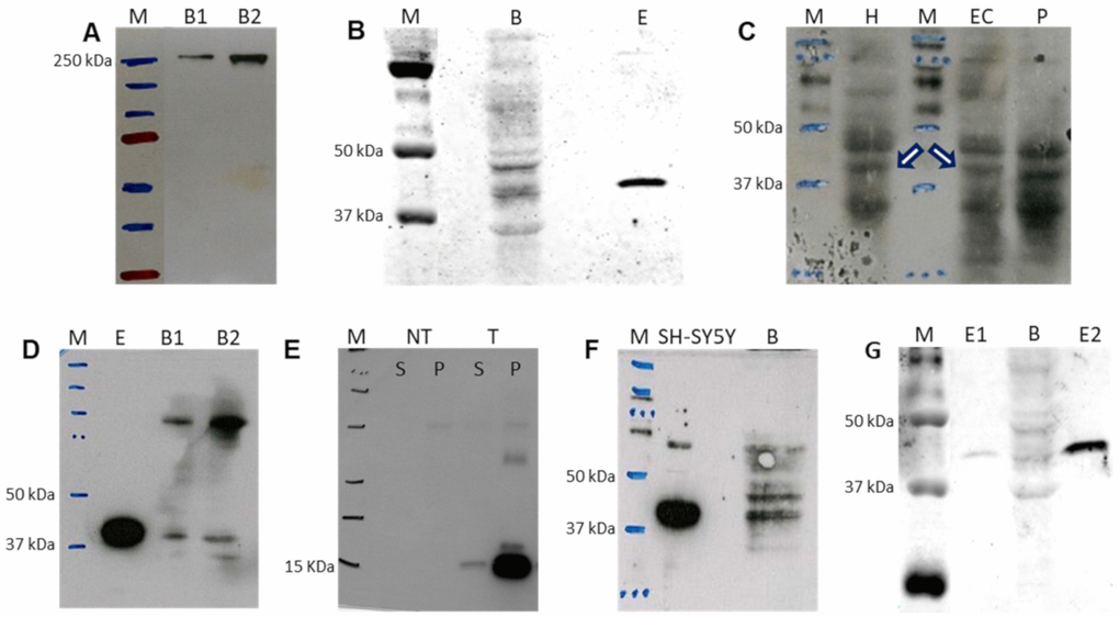
Figure 6.Western blot detection of selected proteins from the de novo pyrimidine biosynthesis pathway in adult human brain. (A) CAD protein in 40 and 80 μg of brain sample, B1 and B2, respectively. (B) DHODH protein in 180 μg of brain (B) protein using polyclonal antibody. Lane E: commercial DHODH enzyme lacking its 31 first amino acids (250 ng of protein). (C) DHODH protein in hippocampus (H, 100 μg of protein), entorhinal cortex (EC, 180 μg of protein) and putamen (P, 180 μg of protein) using polyclonal antibody. White arrows indicate the corresponding band for DHODH. (D) DHODH protein using monoclonal antibody for detection of commercial enzyme lacking its first 31 amino acids (E, 250 ng of protein) and in 40 and 80 μg of brain sample, B1 and B2, respectively. (E) Fragment of DHODH protein used as immunogen to produce the monoclonal antibody. NT and T homogenates of untransformed bacteria and bacteria transformed with the DHODH fragment sequence, respectively. S and P: supernatant and pellet, respectively. (F) DHODH protein in neuroblastoma SH-SY5Y cell line (70 μg of protein) and brain tissue (B, 250 μg of protein) using polyclonal antibody. (G) Quantification of brain DHODH protein with the polyclonal antibody in brain (B, 180 μg of protein) by comparison with the commercial enzyme lacking its first 31 amino acids at 0.4 and 4.0 ng (E1 and E2), respectively. M: molecular weight marker.