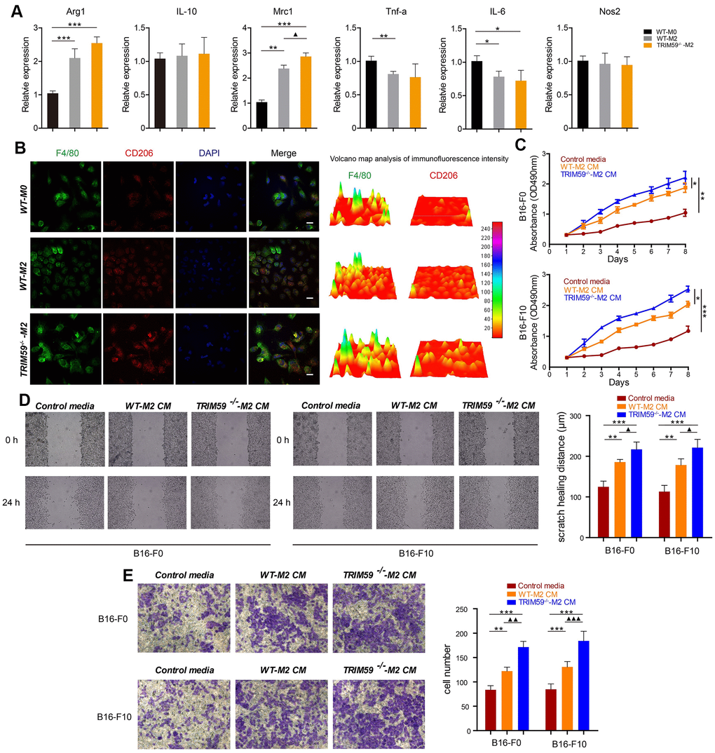
Figure 2.TRIM59-/--M2 macrophage CM promotes melanoma cell migration and invasion. (A) The expression of Arg1, IL-10, Mrc1, TNF-α, IL-6, and NOS2 was detected by qRT-PCR. Data are represented as mean ± SD. *p<0.05, **p<0.01, ***p<0.001, compared with the WT-M0 group; ▲p<0.05, TRIM59-/--M2 vs. WT-M2. (B) Immunofluorescent detection of F4/80 and CD206. Fluorescence intensity was analyzed by a 3D surface plot using ImageJ. Scale bars = 100 μm. (C) Proliferation assay results from B16-F0 and B16-F10 cells treated with control media, WT-M2 CM, or TRIM59-/--M2 CM. Data are represented as mean ± SD. *p<0.05, **p<0.01, ***p<0.001, compared withTRIM59-/--M2 CM. (D) Representative images from wound healing (cell migration) assays and data quantification. Data are represented as mean ± SD. **p<0.01, ***p<0.001, compared with control media; ▲p<0.05, TRIM59-/--M2 CM vs. WT-M2 CM. (E) Representative images from transwell (cell invasion) assays and data quantification. Data are represented as mean ± SD. **p<0.01, ***p<0.001, compared with control media; ▲▲p<0.01, ▲▲▲p<0.001, TRIM59-/--M2 CM vs. WT-M2 CM.